. 2020 Nov 24;117(47):29883-29893.
doi: 10.1073/pnas.2018960117. Epub 2020 Nov 9.

Neuropsychological and neuropathological observations of a long-studied case of memory impairment

Affiliations

Neuropsychological and neuropathological observations of a long-studied case of memory impairment

Larry R Squire et al. Proc Natl Acad Sci U S A. .

Abstract

We report neuropsychological and neuropathological findings for a patient (A.B.), who developed memory impairment after a cardiac arrest at age 39. A.B. was a clinical psychologist who, although unable to return to work, was an active participant in our neuropsychological studies for 24 y. He exhibited a moderately severe and circumscribed impairment in the formation of long-term, declarative memory (anterograde amnesia), together with temporally graded retrograde amnesia covering ∼5 y prior to the cardiac arrest. More remote memory for both facts and autobiographical events was intact. His neuropathology was extensive and involved the medial temporal lobe, the diencephalon, cerebral cortex, basal ganglia, and cerebellum. In the hippocampal formation, there was substantial cell loss in the CA1 and CA3 fields, the hilus of the dentate gyrus (with sparing of granule cells), and the entorhinal cortex. There was also cell loss in the CA2 field, but some remnants remained. The amygdala demonstrated substantial neuronal loss, particularly in its deep nuclei. In the thalamus, there was damage and atrophy of the anterior nuclear complex, the mediodorsal nucleus, and the pulvinar. There was also loss of cells in the medial and lateral mammillary nuclei in the hypothalamus. We suggest that the neuropathology resulted from two separate factors: the initial cardiac arrest (and respiratory distress) and the recurrent seizures that followed, which led to additional damage characteristic of temporal lobe epilepsy.

Keywords: amnesia; diencephalon; hippocampus.


Conflict of interest statement

The authors declare no competing interest.

Figures

Fig. 1.
Fig. 1.
Participants were instructed to copy the figure (67) at the Top Left and 5 to 10 min later to reproduce it from memory. Copies (Top) and reproductions (Bottom) are shown for A.B., for another memory-impaired patient (L.M.) (14), and for a representative control (CON). Although the patients could copy the figure accurately (copy scores: A.B. = 32; L.M. = 33; CON = 31; maximum = 36), they were impaired at reproducing it from memory (reproduction scores: A.B. = 4; L.M. = 6; CON = 26).
Fig. 2.
Fig. 2.
Retrograde memory. (A) Recall performance on a test of news events that occurred from 1952 to 2005 (data from ref. 40). The scores for A.B. show his performance for the period of anterograde amnesia (AA, 1977 to 2005) and for 5-y intervals preceding the onset of amnesia. The scores for controls show performance for the corresponding time periods. (B) Number of details contained in remote autobiographical recollections by A.B. and controls (CON, n = 10) (42). A.B. was given cue words (e.g., river, bird, nail) and tried to recollect an event from early life related to each word. Narratives were scored separately for details that described a specific event (episodic) and for details that were part of the narrative but were not unique to the event (semantic). Brackets show SEM.
Fig. 3.
Fig. 3.
Performance of A.B. and controls (CON, n = 7) on 12 tests of perceptual identification priming and six parallel tests of recognition memory (45). Priming was scored as percent correct identification of 24 study words, presented briefly at test, minus percent correct identification of briefly presented 24 new words. Two-alternative, forced-choice recognition was scored as mean percent correct on six tests that presented 24 pairs of old and new words. Brackets indicate SEM.
Fig. 4.
Fig. 4.
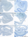
Coronal, Nissl-stained sections of the rostral, anterior, medial temporal lobe arranged from rostral (A) to caudal (C). All images are from the right hemisphere. (Left) Images from the brain of A.B. (Right) Images from the control comparison brain (IML-13) at approximately the same level. (A and A′) These sections illustrate a level through the rostral amygdala (Amyg) and demonstrate cell loss throughout. The entorhinal cortex (EC), located ventromedially, demonstrates patchy cell loss in A.B., particularly in layer III. (B and B′) This section is at a level through the caudal amygdala and at the rostral-most portion of the hippocampus. In the control section (B), the lateral (L), basal (B), and accessory basal (AB) nuclei are easily differentiable. In A.B., there is generalized neuronal loss in the amygdala (B) which is replaced by a background of gliosis. The layers of the entorhinal cortex are apparent in A.B. but are overall less distinct due to patchy cell loss. (C and C’) This section is at the uncal level of the hippocampal formation and illustrates the rostral dentate gyrus (DG), hippocampus (CA3 and CA1), and components of the subicular complex (subiculum [S] and presubiculum [PrS]). Neurons of the polymorphic layer of the dentate gyrus are completely missing at this level as is the CA3 field of the hippocampus (asterisk between the two arrows in C). There is also neuronal loss in the deep portion of the CA1 pyramidal cell layer, which is better seen in Fig. 6).
Fig. 5.
Fig. 5.
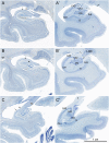
Coronal Nissl-stained sections of the medial temporal lobe of the right hemisphere arranged from rostral (A) to caudal (C). (Left) Images from the brain of A.B. (Right) Images from the control comparison brain (IML-13) at approximately the same level. The major loss of neurons in the sections from A.B. was in the CA3 region of the hippocampus (asterisk and up to arrow) and in the polymorphic layer of the dentate gyrus. (A and A′) Images of the hippocampal formation just caudal to the uncus. There are scattered cells at the arrow in a region containing the CA2 portion of the hippocampus. There is also cell loss in the deep portion of the pyramidal cell layer in the CA1 region of the hippocampus (better seen in Fig. 6). Some patchy cell loss is also observed in the subiculum (S). The presubiculum has a normal appearance. The parahippocampal region, like many other cortical areas, appears to be thinner than usual with patches of cell loss. (B and B′) Images of the hippocampal formation at the level of the caudal lateral geniculate nucleus (LGN). The pathology is similar to what was described in A. An expanded space in the region of the optic radiations (or) may correspond to the damage identified in an early CT scan of A.B. It may also be the cause of the patchy appearance of the cortex in V1 and adjacent visual areas (Fig. 9F) (C and C′) Images of the caudal pole of the hippocampal formation as the fimbria/fornix (f) ascends. The CA3 field of the hippocampus is completely devoid of pyramidal neurons (asterisk), and the polymorphic layer of the dentate gyrus is also totally depopulated of neurons.
Fig. 6.
Fig. 6.
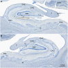
Higher magnification images of the right (A) and left (B) hippocampal formation in A.B. These sections are taken at a level through the rostral lateral geniculate nucleus (LGN). Here, it is clear that the granule cell layer (gl) of the dentate gyrus (DG) is irregular. Much of the polymorphic layer (pl) of the dentate gyrus is depopulated of neurons, but there are some scattered cells in the medial portion of the polymorphic layer (arrows). There appear to be remaining neurons in the CA2 field of the hippocampus (arrow with white arrowhead). It is also easy to appreciate that the deep portion of the CA1 pyramidal cell layer (asterisks) is devoid of neurons. The subiculum (S) and presubiculum (PrS) have an essentially normal appearance with occasional patches of cell loss. Additional abbreviation: ml, molecular layer of the dentate gyrus.
Fig. 7.
Fig. 7.
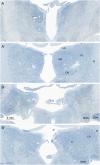
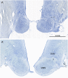
Nissl-stained images of the thalamus. (A and A′) This section is at a mid-rostrocaudal level with A showing A.B. and A′ showing a similar level from the control brain. There is substantial disruption of the structure of the thalamus in A.B. At this level, the mediodorsal (MD) nucleus is nearly entirely depopulated of neurons, and areas of gliosis (outlined region) are extensive. The ventral lateral (VL) nucleus located lateral to the mediodorsal nucleus is somewhat better preserved. The reticular nucleus (R) that surrounds the main portion of the thalamus cannot be distinguished in the section from A.B. (B and B′) These sections are taken through a caudal level of the thalamus that includes the pulvinar (P) and lateral posterior (LP) nuclei. The medial geniculate nucleus (MGN) can also be seen at this level. Virtually all of the pulvinar in A.B. is devoid of neurons, and there are large gliotic patches (outlined area). The medial (MGN) and lateral (LGN) geniculate nuclei of the thalamus are present in the section from A.B. and have a normal appearance. Additional abbreviations: CM, centre median nucleus of the thalamus; LD, lateral dorsal nucleus of the thalamus.
Fig. 8.
Fig. 8.
Nissl-stained sections from A.B. (A) and the control (A′) at a level through the main portion of the medial mammillary nucleus (MMN). In the control brain (A′), there are numerous neurons in the MMN. These are surrounded by a capsule of fibers that include a contribution from the incoming fornix (f) and collecting fibers that will contribute to the mammillothalamic tract. There are also larger and more darkly stained neurons located dorsal to the MMN that constitute the supramammillary region (SUM). In the section from A.B., the MMN is shrunken, and neurons are not detectable. The fibrous capsule is not obvious, and what appears to be the fornix is mainly gliotic. The neurons of the SUM are apparent but appear to be denser due to shrinkage of the lateral hypothalamic tissue. The arrow points to the shrunken fimbria in A.
Fig. 9.
Fig. 9.
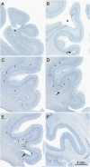
Images of Nissl-stained sections showing neuropathology in the cerebral cortex. (A) Infarct (*) in the left dorsomedial cortex. (B) Infarct (*) in the left posterior parietal cortex. (CE) Sections arranged from rostral (C) through caudal (E) through the right cingulate cortex. Arrows point to patches of cell loss that occurred in all layers and was particularly consistent (and bilateral) in the cingulate cortex. Large arrows in panels B, C, and D point to expanded blood vessels in the white matter. (F) Radially oriented and laminar patches of cell loss in V1 cortex along both banks of the calcarine sulcus. The radial appearance of much of this pathology resembles ocular dominance columns. This may be due to damage to the optic radiations, which is observable in the right hemisphere (Fig. 5B).

Similar articles

Cited by

References

    1. Winslow F., On Obscure Diseases of the Brain and Disorders of the Mind (John W. Davies, London, ed. 2, 1861).
    1. Ribot T., Les Maladies de la Memoire (Appleton-Century-Crofts, New York, 1881) [English translation: Diseases of Memory].
    1. Whitty C. W. M., Zangwill O. L., Amnesia (Butterworths, London, 1966), p. 217.
    1. Milner B., Disorders of learning and memory after temporal lobe lesions in man. Clin. Neurosurg. 19, 421–466 (1972). - PubMed
    1. Squire L. R., The neuropsychology of human memory. Annu. Rev. Neurosci. 5, 241–273 (1982). - PubMed

Publication types

MeSH terms