

Optical coherence tomography in the 2020s-outside the eye clinic
- PMID: 33168975
- PMCID: PMC7853067
- DOI: 10.1038/s41433-020-01263-6
Optical coherence tomography in the 2020s-outside the eye clinic
Abstract
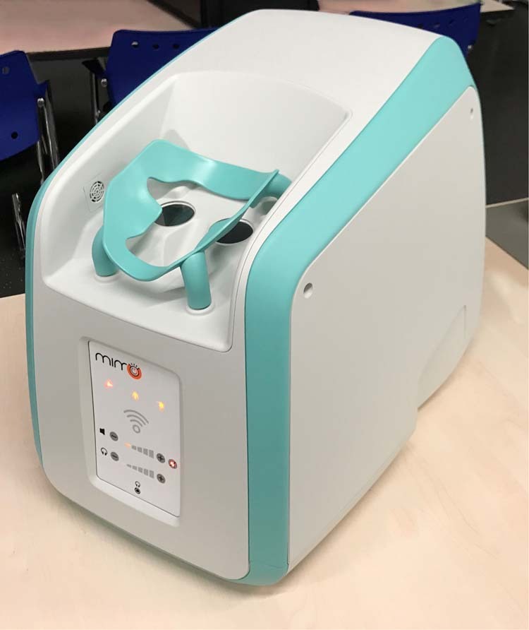
Optical coherence tomography (OCT) is a paragon of success in the translation of biophotonics science to clinical practice. OCT systems have become ubiquitous in eye clinics but access beyond this is limited by their cost, size and the skill required to operate the devices. Remarkable progress has been made in the development of OCT technology to improve the speed of acquisition, the quality of images and into functional extensions of OCT such as OCT angiography. However, more needs to be done to radically improve the access to OCT by addressing its limitations and enable penetration outside of typical clinical settings and into underserved populations. Beyond high-income countries, there are 6.5 billion people with similar eye-care needs, which cannot be met by the current generation of bulky, expensive and complex OCT systems. In addition, advancing the portability of this technology to address opportunities in point-of-care diagnostics, telemedicine and remote monitoring may aid development of personalised medicine. In this review, we discuss the major milestones in OCT hardware development to reach those beyond the eye clinic.
摘要: 相干光断层扫描成像技术 (Optical coherence tomography, OCT) 是将生物光子学在临床实践中应用的成功典范 。在眼科门诊中, OCT 系统得到了广泛应用, 但是由于其成本高、体积大以及需要具备操作技能的原因, OCT 系统在门诊之外的应用受到限制。目前, OCT 技术的发展在提高图像获取速度、图片质量以及OCT在血管功能检测的拓展应用 (如OCT血管成像技术) 方面取得了显著进展。然而, 我们还需通过解决OCT技术的局限性问题以从根本上改善OCT的应用范围方面做更多的工作, 将OCT技术在典型的临床环境之外以及医学资源相对匮乏的地区人群中得以应用。在高收入国家之外, 还有65亿人有类似的眼保健需求, 而目前这一代OCT系统因其庞大、昂贵和复杂而无法满足这些需求。此外, 提高OCT设备的便携性可为医疗点诊断、远程医疗以及远程监护提供可能, 这有助于个体化医疗的发展。在这篇综述中, 我们主要讨论了OCT硬件发展中为实现OCT技术在眼科门诊之外应用目标的里程碑式的成绩。.
Conflict of interest statement
PAK has acted as a consultant for DeepMind, Roche, Novartis and Apellis and is an equity owner in Big Picture Medical. He has received speaker fees from Heidelberg Engineering, Topcon, Allergan and Bayer. RC is an employee of Google LLC and owns Alphabet stock.
Figures

References
-
- Olson J, Sharp P, Goatman K, Prescott G, Scotland G, Fleming A, et al. Improving the economic value of photographic screening for optical coherence tomography-detectable macular oedema: a prospective, multicentre, UK study. Health Technol Assess. 2013;17:1–142.. doi: 10.3310/hta17510. - DOI - PMC - PubMed
-
- Food and Drug Administration. 510(k) Premarket notification, Envisu Spectral Domain Ophthalmic Imaging System (SDOIS). 2012. https://www.accessdata.fda.gov/scripts/cdrh/cfdocs/cfPMN/pmn.cfm?ID=K120057. Accessed 23 Aug 2020.
Publication types
MeSH terms
Grants and funding
LinkOut - more resources
Full Text Sources
Miscellaneous
