

Review of retinal cameras for global coverage of diabetic retinopathy screening
- PMID: 33168977
- PMCID: PMC7852572
- DOI: 10.1038/s41433-020-01262-7
Review of retinal cameras for global coverage of diabetic retinopathy screening
Abstract
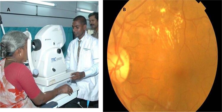
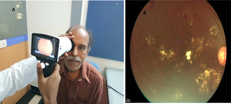
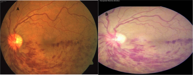
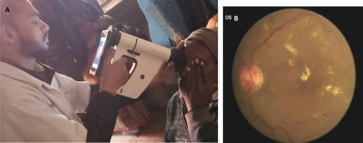
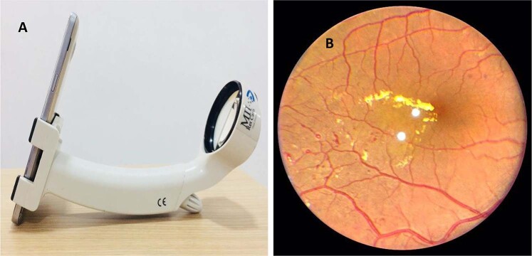
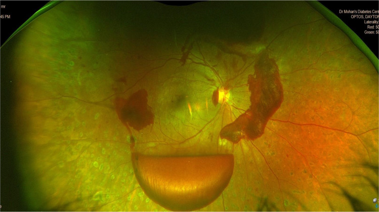
The global burden of diabetes has resulted in an increase in the prevalence of diabetic retinopathy (DR), a microvascular complication of diabetes. Lifelong repetitive screening for DR is essential for early detection and timely management to prevent visual impairment due to the silent sight-threatening disorder. Colour fundus photography (CFP) is helpful for documentation of the retinopathy as well as for counselling the patient. CFP has established roles in DR screening, detection, progression and monitoring of treatment response. DR screening programmes use validated mydriatic or non-mydriatic fundus cameras for retinal imaging and trained image graders identify referable DR. Smartphone-based fundus cameras and handheld fundus cameras that are cost-effective, portable and easy to handle in remote places are gaining popularity in recent years. The images captured with these low-cost devices can be immediately sent to trained ophthalmologists for grading of DR. Recent increase in numbers of telemedicine programmes based on imaging with digital fundus cameras and remote interpretation has facilitated larger population coverage of DR screening and timely referral of those with sight-threatening DR to ophthalmologists. Good-quality retinal imaging and accurate diagnosis are essential to reduce inappropriate referrals. Advances in digital imaging such as ultra-wide field imaging and multi-modal imaging have opened new avenues for assessing DR. Fundus cameras with integrated artificial intelligence (AI)-based automated algorithms can also provide instant DR diagnosis and reduce the burden of healthcare systems. We review the different types of fundus cameras currently used in DR screening and management around the world.
摘要: 在全球, 糖尿病的患病率日益增长导致了微血管并发症—糖尿病视网膜病变(DR)的患病率增加。为了早期发现和及时治疗, 对DR患者的终身反复筛查至关重要, 以防因这种隐匿的视力威胁造成视力损害。彩色眼底照相(CFP)有助于记录视网膜病变以及有利于患者进行咨询。CFP已在DR的筛查、检测、进展和监测治疗反应等方面发挥了重要作用。DR筛查项目使用经过验证的散瞳或非散瞳眼底相机进行视网膜成像并且由训练有素的图像分级员识别DR。智能手机近年来在偏远地区越来越受欢迎, 基于手机的眼底相机和手持式眼底相机具有成本效益低、便携和易于操作的特点。使用这些低成本设备捕获的图像可以立即发送给训练有素的眼科医生, 以便对DR进行分级。最近, 基于数字眼底相机成像和远程解读的远程医疗计划数量增加, 有助于扩大DR筛查的人群覆盖面, 并及时将患有视力损伤的DR患者转诊给眼科医生。高质量的视网膜成像和精准的诊断对于减少不当的转诊至关重要。数字成像的进步, 如超宽范围成像和多模式成像, 为评估DR开辟了新的途径。带有集成人工智能(AI)自动化算法的眼底相机还可以提供即时的DR诊断, 并减轻医疗系统的负担。我们对目前世界各地用于DR筛查和管理的不同类型的眼底相机进行了综述。.
Conflict of interest statement
The authors declare that they have no conflict of interest.
Figures
References
-
- International Diabetes Federation. Diabetes Atlas, 9th ed. Brussels, Belgium: International Diabetes Federation; 2019. http://www.diabetesatlas.org. Accessed 14 Jun 2020.
-
- Flaxman SR, Bourne RRA, Resnikoff S, Ackland P, BraithwaiteT, Cicinelli MV, et al. Vision loss expert group of the Global Burden of Disease Study. Global causes of blindness and distance vision impairment 1990–2020: a systematic review and meta-analysis. Lancet Glob Health. 2017;5:e1221–34. doi: 10.1016/S2214-109X(17)30393-5. - DOI - PubMed
Publication types
MeSH terms
LinkOut - more resources
Full Text Sources
Medical
Miscellaneous
