. 2021 Apr;20(4):560-569.
doi: 10.1038/s41563-020-00844-w. Epub 2020 Nov 9.

Activating an adaptive immune response from a hydrogel scaffold imparts regenerative wound healing

Affiliations

Activating an adaptive immune response from a hydrogel scaffold imparts regenerative wound healing

Donald R Griffin et al. Nat Mater. 2021 Apr.

Abstract

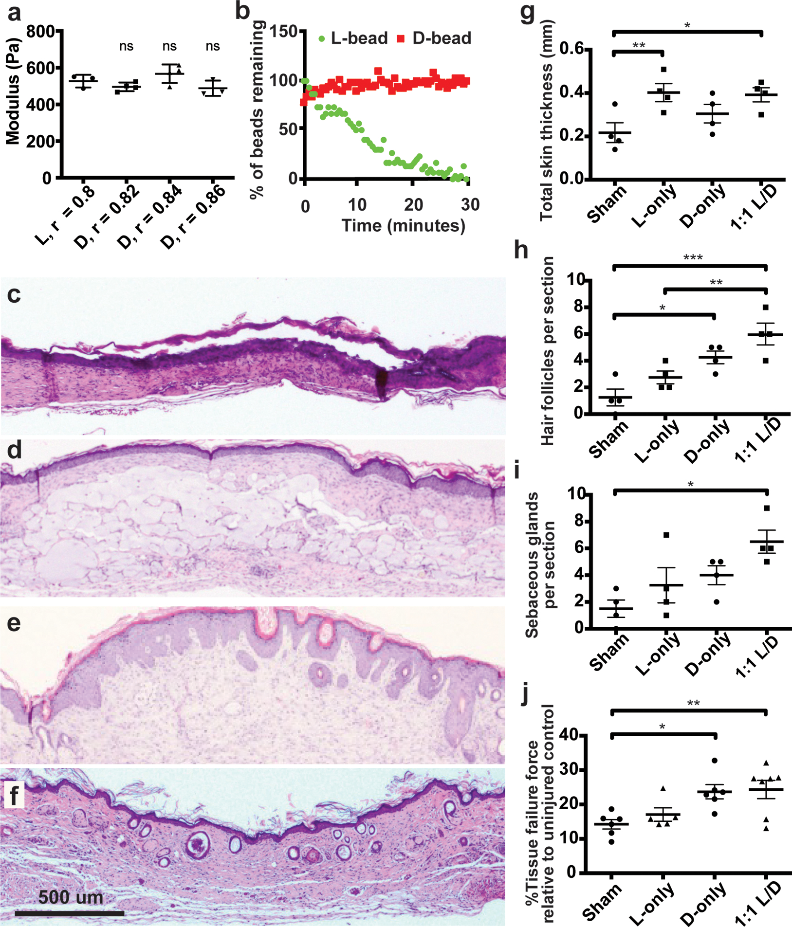
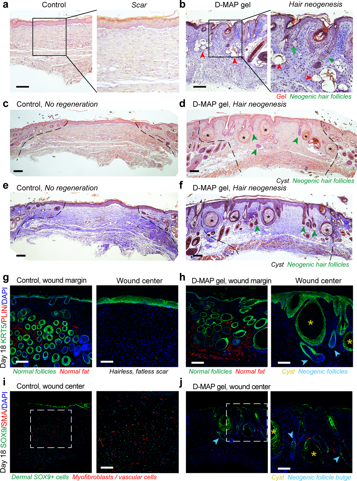
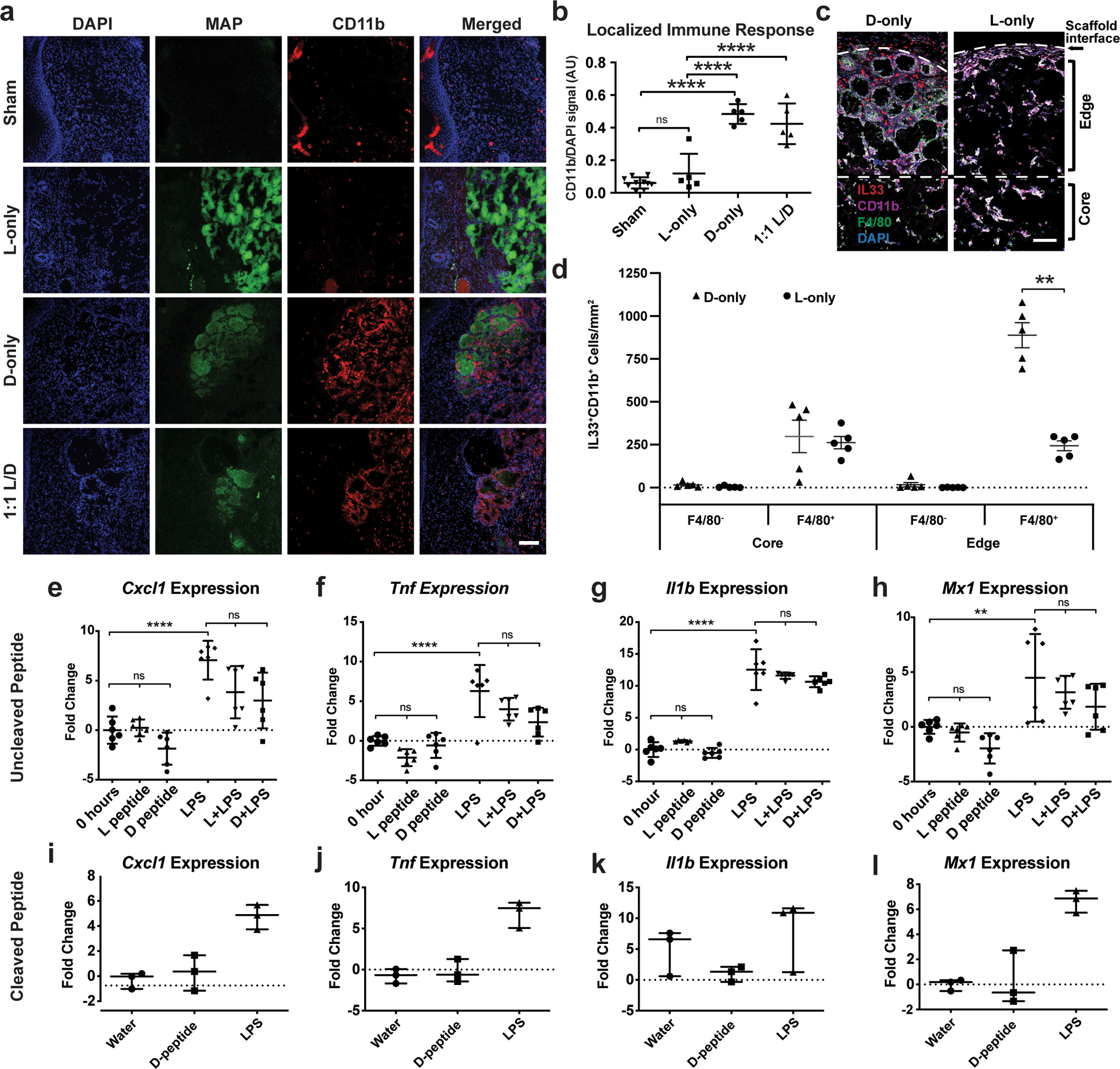
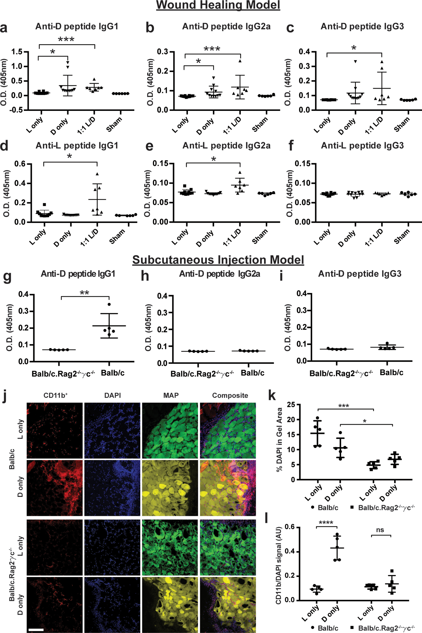
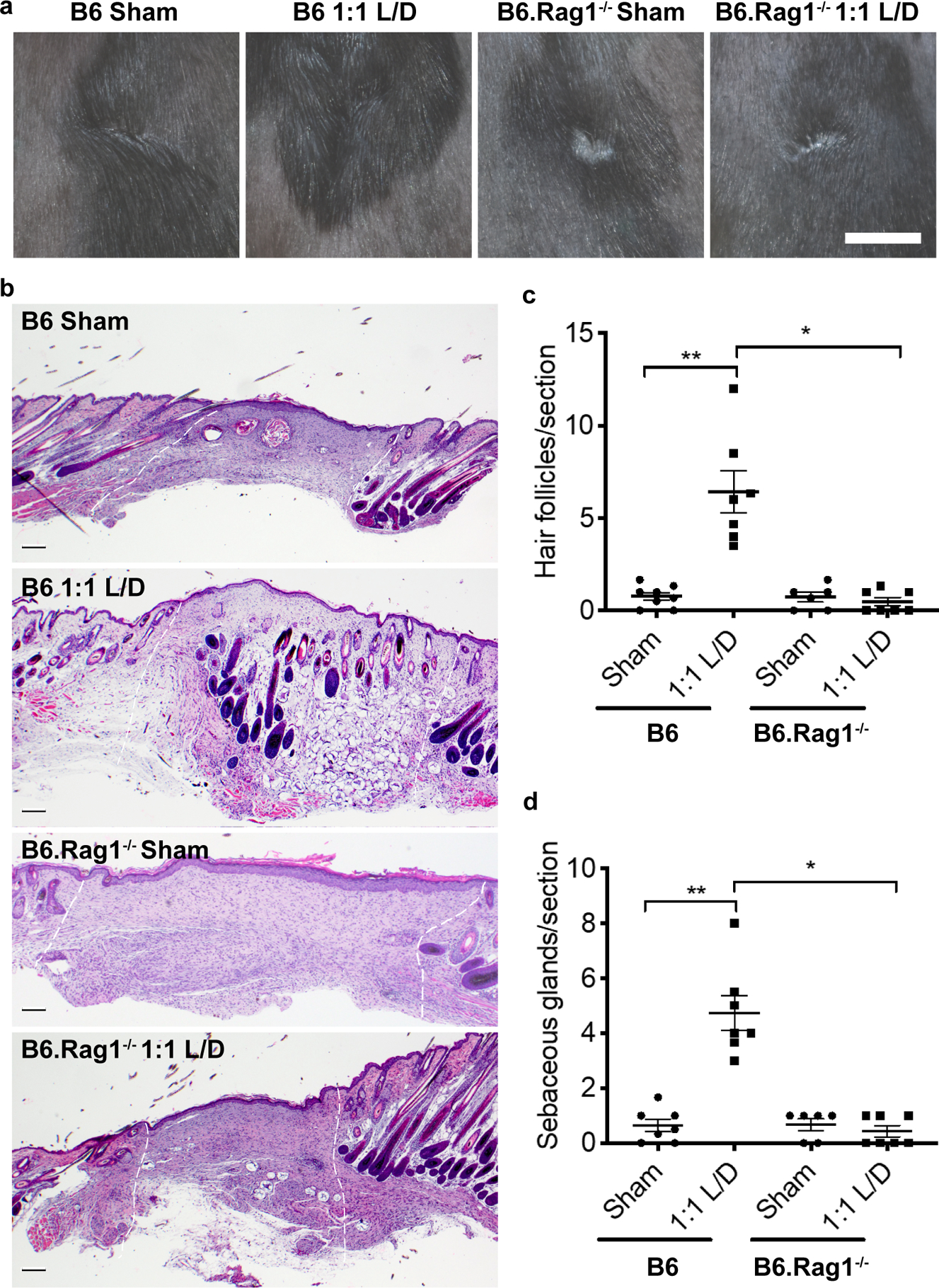
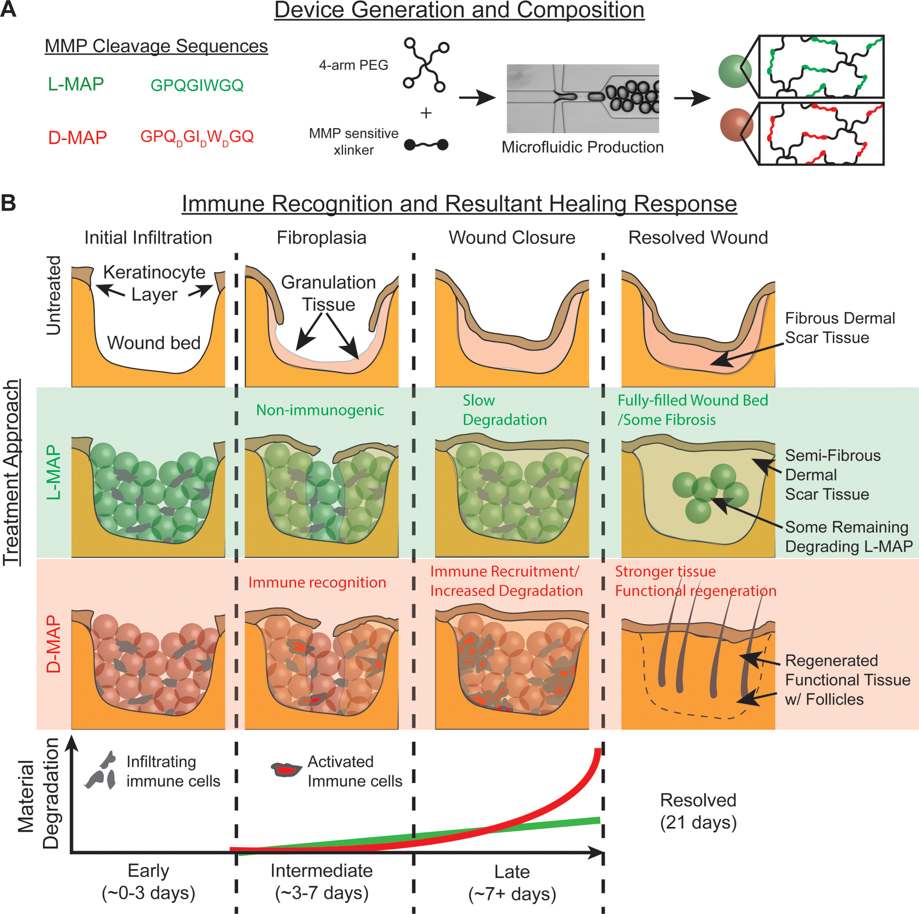
Microporous annealed particle (MAP) scaffolds are flowable, in situ crosslinked, microporous scaffolds composed of microgel building blocks and were previously shown to accelerate wound healing. To promote more extensive tissue ingrowth before scaffold degradation, we aimed to slow MAP degradation by switching the chirality of the crosslinking peptides from L- to D-amino acids. Unexpectedly, despite showing the predicted slower enzymatic degradation in vitro, D-peptide crosslinked MAP hydrogel (D-MAP) hastened material degradation in vivo and imparted significant tissue regeneration to healed cutaneous wounds, including increased tensile strength and hair neogenesis. MAP scaffolds recruit IL-33 type 2 myeloid cells, which is amplified in the presence of D-peptides. Remarkably, D-MAP elicited significant antigen-specific immunity against the D-chiral peptides, and an intact adaptive immune system was required for the hydrogel-induced skin regeneration. These findings demonstrate that the generation of an adaptive immune response from a biomaterial is sufficient to induce cutaneous regenerative healing despite faster scaffold degradation.


Conflict of interest statement

Competing financial interests

D.R.G., W.M.W., D.D.C., T.S., and P.O.S. have a financial interest in Tempo Therapeutics, which aims to commercialize MAP technology.

Figures

Figure 1.
Figure 1.. D-MAP hydrogel degradation is enhanced in wounds of SKH1 hairless mice.
a) Rheological characterization of MAP hydrogels composed of L or D-peptide crosslinked microgels. The r-ratio (ratio of -SH to -VS) used to form the microgels was changed to arrive at the same storage modulus for both L and D MAP scaffolds. NS represents a no statistical significance between the L MAP scaffold to the D-MAP scaffold indicated using a two-tailed student t-test. b) Fabricated L or D hydrogels were tested for in vitro enzymolysis behavior through exposure to a solution of collagenase I (5U/mL). c-f) Representative low power view of H&E sections from healed skin 21 days after splinted excisional wounding from a Sham (c), L-MAP (d), D-MAP (e), and 1:1 mixture of L-MAP and D-MAP treated wound in SKH1 mice (f). g-i) Histologic quantification of dermal thickness including gels (in mm), hair follicles, and sebaceous glands. Each point represents average of 2 sections from 2 separate slides of one wound. Each data point represents one animal and all analysis is by one-way ANOVA (respective F-values (3,12): 4.448, 10.89, 5.074, stars denote statistical significance by Tukey multiple comparisons test: g) *p=0.0460, **p=0.0341, h) *p=0.0220, **p= 0.0133, ***p=0.0007 i) *p=0.0110). j) 28 days after incisional, unsplinted wounds were created, healed wounds that were treated without or with different hydrogels were tested against unwounded skin in the same mouse. Tensile strength was evaluated by tensiometry and reported as a percentage of the tensile strength of the scar tissue when compared to the normal skin of the same mouse. Each data point represents average of two measurement from one wound, separate from wounds used in b-i with analysis by one-way ANOVA (F-value (3, 20): 5.400, *p=0.0273, **p=0.0131). Data is plotted as a scatter plot showing the mean and standard deviation.
Figure 2:
Figure 2:. D-MAP hydrogel induces neogenesis of hair follicles in full-thickness skin wounds in B6 mice.
a-f) H&E (a, c, d) and Trichrome staining (b, e, f) of healed 4-mm full-thickness splinted skin wound on day 18. Control (sham-treated) wounds heal with scarring (a, c, e), while D-MAP gel treated wounds form numerous epidermal cysts (asterisks) and, prominently, regenerate de novo hair follicles (green arrowheads) (b, d, f). In some instances, neogenic hair follicles form in close association with epidermal cysts. As compared to normal, pre-existing anagen hair follicles at the wound edges, neogenic hair follicles display early anagen stage morphology (Wound edges in b-d are outlined and D-MAP hydrogel remnants in b are marked with red arrowheads). g-h) Immunostaining for epithelial marker KRT5 (green) and adipocyte marker PLIN (red), reveals normal KRT5+ anagen hair follicles and many mature PLIN+ dermal adipocytes (left panels in g and h). Regeneration of new KRT5+ hair follicles (arrowheads in h) along with KRT5+ epidermal cysts is observed only in D-MAP hydrogel-treated wounds (right panel g vs. h). No neogenic adipocytes are observed in hair-forming D-MAP-treated wounds. i-j) Immunostaining for SOX9 (green) and SMA (red), reveals many SOX9+ epithelial cells within the bulge region of neogenic hair follicles in day 18 DMAP-treated wounds (arrowheads in j). In contrast, in control (sham-treated) wounds that undergo scarring, dermal wound portion contains many Sox9+ cells, many of which also co-express contractile marker SMA (i). Expression of SMA is also seen in both control and D-MAP-treated samples in blood vessels. Scales in a-j = 100 μm. The images are representative of slides from 4 animals per group.
Figure 3.
Figure 3.. Peptide recognition by pattern recognition receptors is not required for myeloid cell recruitment.
a) Representative confocal immunofluorescent images staining myeloid cells (CD11b+) within healed wounds of B6 mice in the absence or presence of the indicated hydrogel. Scale = 100μm. b) Quantification of CD11b+ cellular infiltrate in healed tissue 21 days after wounding in the presence or absence of hydrogel. Each point represents average of 3 slides for each wound. All analysis is by one-way ANOVA (F-value (3,21): 41.10; **** denotes p<0.0001). c-d) Representative high-resolution confocal immunofluorescence imaging for CD11b, F4/80, DAPI, and IL-33 from subcutaneous implants of L- or D-MAP hydrogel implants (c) and quantification of IL-33 producing macrophages and other myeloid cells at hydrogel edge and core. n=5 B6 mice, mean +/− SEM (d), multiple t-tests adjusted for multiple comparisons using Holm-Sidak method (** denotes p=0.00014). Scale = 100μm. e-h) Murine bone marrow derived macrophages (BMDMs) from B6 mice were stimulated with 500 μg/ml of full-length L- or D- crosslinker peptide in the presence or absence of lipopolysaccharide (10ng/ml) for 6 hours. Shown are qPCR results of 4 inflammatory genes from two separate experiment performed with n = 6. All analysis is by one-way ANOVA (Respective F-values (5, 30): 15.66, 17.62, 107.1, and 8.229, ** denotes p=0.009 and **** denotes p<0.0001) i-l) BMDMs were stimulated with LPS (10ng/ml) or cleaved D-crosslinker peptide (500μg/ml) that possessed an N-terminal D-amino acid. Experiment was performed in triplicate. All analysis is by one-way ANOVA (Respective F-values (2,6): 20.28, 30.86, 2.178, and 22.72). Data is plotted as a scatter plot showing the mean and standard deviation.
Figure 4.
Figure 4.. D-MAP induces antibody responses and recruitment of myeloid cells via adaptive immunity.
a-c) Measurement of anti-D specific IgG subtype antibodies by ELISA 21 days following wound healing experiments in SKH1 mice treated with indicated hydrogels. d-f) Measurement of anti-L specific IgG subtype antibodies by ELISA 21 days following wound healing experiments in SKH1 mice treated with indicated hydrogels. Each data point represents one animal and all analysis in a-f is by unpaired two-tailed t-test comparing each condition to L only. g-i) Measurement of anti-D specific IgG subtype antibodies in Balb/c or Balb/c.Rag2−/−γc−/− mice given a subcutaneous injection of D-MAP 21 days after injection. Each data point represents one animal and all analysis in g-I is by unpaired two-tailed t-test (** denotes p=0.0022). j-l) Representative examples of confocal immunofluorescent imaging for CD11b, DAPI, and hydrogel from subcutaneous implants of L- or D-MAP hydrogel implants in Balb/c or Balb/c.Rag2−/−γc−/− mice. Scale = 200μm. (j) and quantification of total DAPI+ cells (k) and CD11b+ myeloid cells (l). Data is plotted as a scatter plot showing the mean and standard deviation. Each point represents average of 3 slides for each wound. All analysis is by unpaired two-tailed t-test (* denotes p=0.0455, *** denotes p=0.0006, **** denotes p<0.0001) represent statistical significance by student t-test for the comparison indicated.
Figure 5.
Figure 5.. D-MAP requires an intact adaptive immunity to induce hair follicle neogenesis.
a) Representative examples of gross clinical images of healed splinted excisional wounds in B6 or B6.Rag1−/− mice by DSLR camera 17 days later treated as sham (no hydrogel) or 1:1 L/D-MAP treatment. Scale = 5mm. b) Histologic sections of healed tissue from B6 or B6.Rag1−/− mice. Scale = 200μm. White dashed lines denotes wounded area. Quantification of the average numbers of c) hair follicles and d) sebaceous glands from 3 histological sections per sample from B6 mice and B6.Rag1−/− mice. Data is plotted as a scatter plot showing the mean and SEM. * denotes two-tailed p=0.002 by Mann Whitney test, for inter-strain/identical treatment comparison and ** denotes p=0.0039 by Wilcoxon test for intra-strain/different treatment comparison.
Figure 6.
Figure 6.. D-MAP changes the wound fate from scar formation to regeneration by type 2 immune activation.
a) Representation of amino acid chirality within the cross-linking peptides, microfluidic formation of the hydrogel microbeads incorporating L- or D- chirality peptides. b) The use of L- or D-MAP in a wound healing model demonstrates that both L- or D-MAP hydrogel fill the wound defect. While wounds that heal in the absence of hydrogel heal with an atrophic scar with loss of tissue, the epidermis forms over the scaffold in both cases and allows for increased dermal thickness. However, in the case of D-MAP, the hydrogel activates the adaptive immune system over time, resulting in tissue remodeling and skin regeneration as the adaptive immune system degrades the D-MAP scaffold.

Comment in

  • MAP-ing a way towards tissue repair.
    Stelzel JL, Doloff JC. Stelzel JL, et al. Nat Mater. 2021 Apr;20(4):452-453. doi: 10.1038/s41563-021-00963-y. Nat Mater. 2021. PMID: 33772230 No abstract available.

References

    1. Griffin DR, Weaver WM, Scumpia P, Di Carlo D & Segura T Accelerated wound healing by injectable microporous gel scaffolds assembled from annealed building blocks. Nat. Mater 14, 737–744 (2015). - PMC - PubMed
    1. Nih LR, Sideris E, Carmichael ST & Segura T Injection of Microporous Annealing Particle (MAP) Hydrogels in the Stroke Cavity Reduces Gliosis and Inflammation and Promotes NPC Migration to the Lesion. Adv. Mater. Deerfield Beach Fla 29, (2017). - PMC - PubMed
    1. Xu Q et al. Injectable hyperbranched poly(β-amino ester) hydrogels with on-demand degradation profiles to match wound healing processes. Chem. Sci 9, 2179–2187 (2018). - PMC - PubMed
    1. Zhu S, Nih L, Carmichael ST, Lu Y & Segura T Enzyme-Responsive Delivery of Multiple Proteins with Spatiotemporal Control. Adv. Mater. Deerfield Beach Fla 27, 3620–3625 (2015). - PMC - PubMed
    1. Sela M & Zisman E Different roles of D-amino acids in immune phenomena. FASEB J. Off. Publ. Fed. Am. Soc. Exp. Biol 11, 449–456 (1997). - PubMed

Publication types