Review
. 2021 Jan;24(1):5-18.
doi: 10.1038/s41593-020-00738-9. Epub 2020 Nov 9.

Prefrontal-amygdala circuits in social decision-making

Affiliations
Review

Prefrontal-amygdala circuits in social decision-making

Prabaha Gangopadhyay et al. Nat Neurosci. 2021 Jan.

Abstract

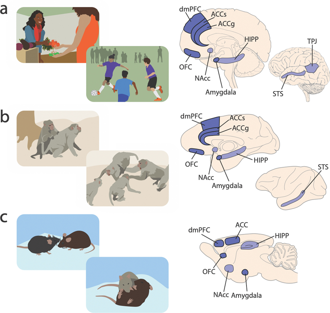
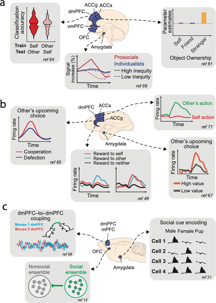
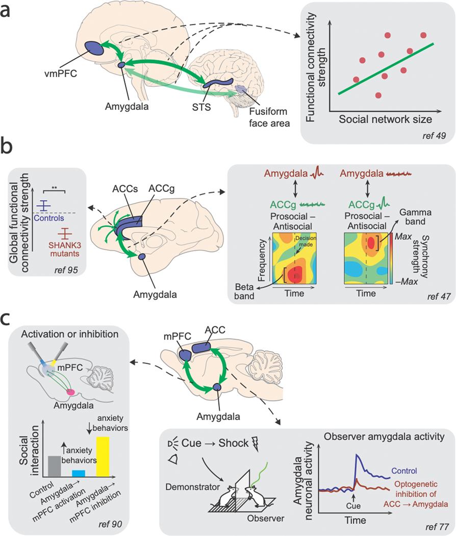
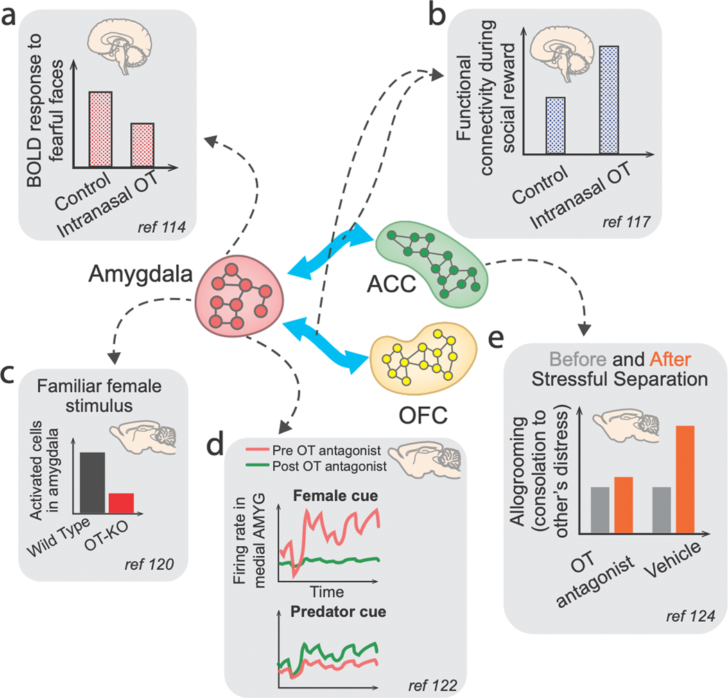
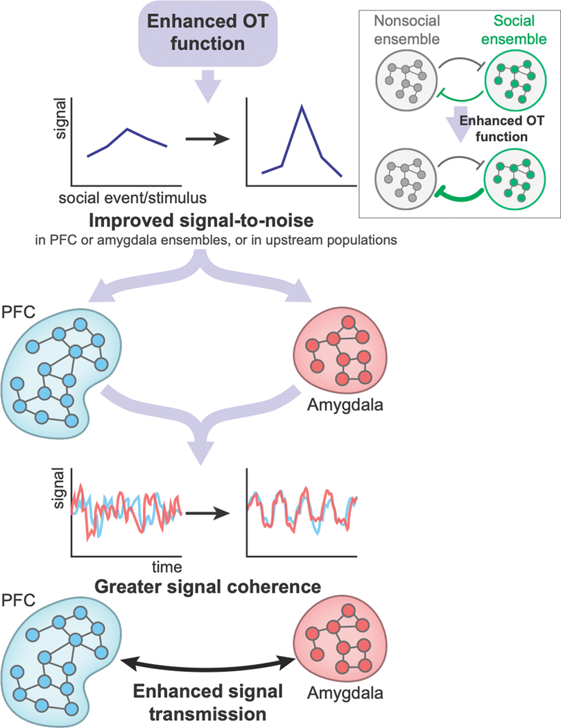
An increasing amount of research effort is being directed toward investigating the neural bases of social cognition from a systems neuroscience perspective. Evidence from multiple animal species is beginning to provide a mechanistic understanding of the substrates of social behaviors at multiple levels of neurobiology, ranging from those underlying high-level social constructs in humans and their more rudimentary underpinnings in monkeys to circuit-level and cell-type-specific instantiations of social behaviors in rodents. Here we review literature examining the neural mechanisms of social decision-making in humans, non-human primates and rodents, focusing on the amygdala and the medial and orbital prefrontal cortical regions and their functional interactions. We also discuss how the neuropeptide oxytocin impacts these circuits and their downstream effects on social behaviors. Overall, we conclude that regulated interactions of neuronal activity in the prefrontal-amygdala pathways critically contribute to social decision-making in the brains of primates and rodents.


Figures

Figure 1.
Figure 1.. Behavioral ecology of social interaction and brain regions commonly recruited by social behaviors in humans, nonhuman primates, and rodents.
Behavioral illustrations in the left column depict selected social interaction scenarios for humans (a), rhesus macaques (b), and mice (c), exhibiting different levels of complexity in social interactions. Brain illustrations on the right column depict key brain regions that are discussed in this review (darker contrast) and other related regions briefly mentioned in connection (lighter contrast) that are implicated in various social behaviors in each species. (ACCg, anterior cingulate gyrus; ACCs, anterior cingulate sulcus; dmPFC, dorsomedial prefrontal cortex; OFC, orbitofrontal cortex; NAcc, nucleus accumbens; STS, superior temporal sulcus; TPJ, temporal parietal junction; HIPP, hippocampus). Social operations in these brain regions are being actively investigated at multiple neurobiological levels across humans, nonhuman primates, and rodents.
Figure 2.
Figure 2.. Illustrations of selected results demonstrating the importance of PFC and amygdala in social behaviors.
Across humans (a), macaques (b), and mice (c), summary diagrams illustrating selected findings from particular studies are shown with corresponding references. (a) (left) In humans, dmPFC BOLD activations tracked subjective value for making decisions for self as well as on behalf of another individual in a shared manner. (middle) BOLD signals in the human amygdala showed different response patterns to high versus low inequity in reward outcomes between self and another individual for prosocial individuals. (right) BOLD activations in ACCg in the human brain signaled objects belonging to strangers but not to self or friends. (b) (left) Neuronal activity in a population of ACC neurons in monkeys was found to predict if a partner monkey was going to cooperate or defect in a prisoner’s dilemma task. (middle) Neuronal activity in a population of ACCg neurons either exclusively signaled conspecific’s reward outcome or signaled the reward outcome of self or other in a comparable fashion. (top right) Many neurons in dmPFC selectively increased activity to encode other’s action in a turn-taking task between two monkeys. (bottom right) Activity of a group of amygdala neurons in monkeys was found to encode conspecific’s upcoming choice when observing other’s value-guided actions. (c) (top left) During social interactions, mice exhibited dmPFC-to-dmPFC neuronal synchrony in behaviorally relevant manners. (bottom left) In the mouse OFC, neuronal ensembles selective for social behavior were shown to inhibit neuronal ensembles selective for nonsocial behavior. (right) Neurons in the mouse amygdala were shown to discriminate social cues.
Figure 3.
Figure 3.. Illustrations of selected results demonstrating the importance of PFC-amygdala interactions in social behaviors.
(a) In humans, resting-state functional connectivity measures between the amygdala and vmPFC, between the amygdala and STS, as well as between the amygdala and the fusiform face area were shown to index individual participants’ social network size. (b) Macaques with SHANK3 mutation, a model system frequently used to study autism spectrum disorder based on disrupted synaptic communication, exhibit abnormal global functional connectivity patterns involving ACC, among other regions. In addition, coherence between spiking activity and LFP signals between ACCg and the basolateral amygdala was enhanced during prosocial decisions compared to antisocial decisions in distinct frequency channels. (c) In mice, optogenetic activation of basolateral amygdala (BLA) neurons innervating mPFC reduce social interaction (but increased anxiety behaviors), whereas inhibiting the same projection neurons enhanced social interaction (but decreased anxiety behaviors). Moreover, ACC input to BLA neurons was found to be necessary for BLA neurons to signal observational fear cues, and optogenetically inhibiting these BLA-projecting ACC neurons prevented observational fear learning in mice.
Figure 4.
Figure 4.. Illustrations of selected findings showing neuromodulation by OT in PFC–amygdala pathways.
(a) Intranasally administered OT in humans was shown to attenuate amygdala BOLD responses to fearful faces and (b) modulate OFC–amygdala and ACC–amygdala functional connectivity strength when perceiving a socially rewarding stimulus (infant laughter). (c) OT function in the medial amygdala is required for social recognition and for sex discrimination of social cues in mice. (d) Blocking OT function in the medial amygdala in male mice reduced the processing of female cues but increased processing of predator cues. (e) In mice, OT processing in ACC is involved in partner-directed grooming behaviors to a conspecific under distress.
Figure 5.
Figure 5.. A hypothesized mechanism by which OT may enhance social functions in the PFC–amygdala pathways.
OT may improve signal-to-noise ratio of neural signals either in amygdala and PFC neural populations or in neural populations upstream to amygdala or PFC. Moreover, when there are mutual inhibition processes between neural ensembles linked to nonsocial behaviors and neural ensembles linked to social behaviors, OT may strengthen the inhibition of nonsocial ensembles by social ensembles (inset). As a result, OT would enhance neural signal transmission and possibly strengthen synchrony across neural ensembles between amygdala and PFC subregions. According to this hypothesis, OT would therefore enhance social functions that critically depend on PFC-amygdala interactions. Note that this mechanism is likely to be a general means by which many types of neuromodulators modulate various cognitive functions in multiple neural circuits.

References

    1. Chang SWC et al. Neuroethology of primate social behavior. Proc. Natl. Acad. Sci. U. S. A. 110 Suppl 2, 10387–10394 (2013). - PMC - PubMed
    1. Ruff CC & Fehr E. The neurobiology of rewards and values in social decision making. Nat. Rev. Neurosci. 15, 549–562 (2014). - PubMed
    1. Giese MA & Rizzolatti G. Neural and computational mechanisms of action processing: interaction between visual and motor representations. Neuron 88, 167–180 (2015). - PubMed
    1. Apps MAJ, Rushworth MFS & Chang SWC The anterior cingulate gyrus and social cognition: tracking the motivation of others. Neuron 90, 692–707 (2016). - PMC - PubMed
    1. Chang SWC An emerging field of primate social neurophysiology: current developments. eNeuro 4, (2017). - PMC - PubMed

Publication types