Review
. 2021 Apr;28(2):409-421.
doi: 10.1007/s10140-020-01866-2. Epub 2020 Nov 9.

MRI of myositis and other urgent muscle-related disorders

Affiliations
Review

MRI of myositis and other urgent muscle-related disorders

Paul L Wasserman et al. Emerg Radiol. 2021 Apr.

Abstract

Myositis has many etiologies, and it can be encountered in the acute or chronic setting. Our goal is to increase the radiologist's knowledge of myositis and other urgent muscle disorders encountered in the emergent or urgent setting. We review the clinical presentation, the MRI appearance, and the complications that can be associated with these entities. Since myositis can affect multiple muscle compartments, we review how to differentiate the compartments of the appendicular skeletal in order to generate reports that relay important anatomic information to the treating physician. Given the poor sensitivity and positive predictive value of the clinical signs and symptoms used to diagnosing acute compartment syndrome, we discuss the potential use of MRI in cases of suspected but clinically equivocal compartment syndrome in the future.

Keywords: Anatomy; Compartment syndrome; MRI; Myositis; Rhabdomyolysis.


Conflict of interest statement

The authors declare that have no conflict of interest.

Figures
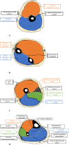
Fig. 1
Fig. 1
Images of anatomic compartments
Fig. 2
Fig. 2
a Stage 2, pyomyositis in a 36-year-old immunosuppressed patient with an irregularly shaped, multiloculated, peripherally enhancing abscess within the posterior compartment of the proximal thigh on this T1 fat-saturated post-contrast image (arrows). Note the enhancement of the surrounding musculature consistent with infectious myositis (star). Axial proton density with fat saturation at the same level b reveals an abnormal area of increased signal intensity that corresponds to the abscess; however, is less well defined (arrows). Increased signal is also noted in the adjacent muscle (star)
Fig. 3
Fig. 3
Viral myositis (HIV) 48-year-old. Axial STIR images through the mid-thighs reveals heterogeneous hyperintense signal affecting all compartments
Fig. 4
Fig. 4
ODCS with type 2, rhabdomyolysis in a 34-year-old. Coronal proton density fat saturation image (a) reveals patchy, heterogeneously hyperintense images of the right hip musculature after being “found down” due to OxyContin overdose. Note the bulging piriformis (star), gluteus minimus (diamond), and gluteus medius (arrowhead) muscles and fluid tracking along the proximal iliotibial band. Coronal T1 image (b) of the same patient
Fig. 5
Fig. 5
Rhabdomyolysis in a 23-year-old patient who presented to the emergency department 1 week after smoking synthetic cannabinoids with a creatine kinase of 203,700. Axial proton density fat-saturated image reveals selective but symmetric bilateral hyperintensity of all compartments with relative sparing of the adductor longus muscles (star) and vastus intermedius muscles (arrowhead) groups
Fig. 6
Fig. 6
Diabetic myonecrosis in a 48-year-old patient who presented to the emergency department with acute calf pain and a long history of uncontrolled diabetes, including nephropathy and retinopathy. Sagittal STIR image (a) reveals heterogeneous, hyperintense signal within the superficial posterior compartment (arrow). Axial PD fat saturation images through the calf (b) reveals heterogenous hyperintense T2-weighted signal throughout the lateral gastrocnemius muscle (arrows). Axial T1 image (c) notes isointense effacement of intermuscular fat within the lateral gastrocnemius relative to the surrounding musculature (arrowhead)
Fig. 7
Fig. 7
Polymyositis. Axial T1-weighted image (a) through the mid-thigh reveals mildly increased but isointense bilateral muscle swelling. Axial proton density fat-saturated image (b) through the same level reveals patchy hyperintense signal within all compartments with relatively sparing of some posterior and medial compartment muscles
Fig. 8
Fig. 8
Dermatomyositis in 59-year-old. Axial STIR image of the lower pelvis reveals patient with diffusely increased hyperintensity of the obturator internus (circle) and obturator externus muscles (arrowhead) due to chronic, steroid-resistant dermatomyositis. Patient succumbed to respiratory failure within 1 month of this image
Fig. 9
Fig. 9
A 59-year-old with rheumatoid arthritis and polymyositis. Axial STIR image of the left mid-thigh reveals patchy hyperintense involvement of all three compartments with some muscle sparing in the medial and posterior compartments. Note relative atrophy of the hyperintense muscles
Fig. 10
Fig. 10
Dorsal compartment syndrome of the forearm. Coronal proton density fat-saturation image (a) reveals heterogeneously hyperintense signal affecting the dorsal compartment due to blunt trauma. Axial short TI inversion recovery (STIR) (b) reveals bowing of the superficial deep fascia secondary to the swollen muscles throughout the dorsal compartment (arrows)

References

    1. Andrew N. Pollak M (2014) United States Bone and Joint Initiative: the Burden of Musculoskeletal Disease in the United States (BMUS). http://www.boneandjointburden.org. Accessed 2 Aug 2020
    1. Johnstone AJ, Ball D (2019) Determining ischaemic thresholds through our understanding of cellular metabolism. BTI - Compartment Syndrome: A Guide to Diagnosis and Management [Internet]. Chapter 4. Springer, Cham - PubMed
    1. Taylor DJ. Clinical utility of muscle MR spectroscopy. Semin Musculoskelet Radiol. 2000;4(4):481–502. doi: 10.1055/s-2000-13172. - DOI - PubMed
    1. Sedivy P, Drobny M, Dezortova M, Herynek V, Roztocil K, Cermakova H, Nemcova A, Dubsky M, Hajek M. 31P-MR spectroscopy in patients with mild and serious lower limb ischemia. Int Angiol. 2018;37(4):293–299. doi: 10.23736/s0392-9590.18.03943-3. - DOI - PubMed
    1. Kennedy PA-O, Barnhill E, Gray C, Brown C, van Beek EJR, Roberts N, Greig CA (2020) Magnetic resonance elastography (MRE) shows significant reduction of thigh muscle stiffness in healthy older adults. (2509–2723 (Electronic))GeroScience 42:311–321. 10.1007/s11357-019-00147-2 - PMC - PubMed

MeSH terms