Review
. 2020 Dec;46(12):2436-2449.
doi: 10.1007/s00134-020-06291-0. Epub 2020 Nov 9.

How to ventilate obstructive and asthmatic patients

Affiliations
Review

How to ventilate obstructive and asthmatic patients

Alexandre Demoule et al. Intensive Care Med. 2020 Dec.

Abstract

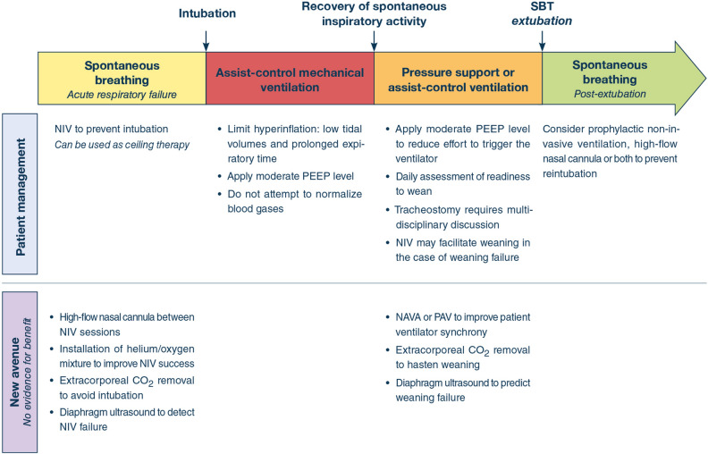
Exacerbations are part of the natural history of chronic obstructive pulmonary disease and asthma. Severe exacerbations can cause acute respiratory failure, which may ultimately require mechanical ventilation. This review summarizes practical ventilator strategies for the management of patients with obstructive airway disease. Such strategies include non-invasive mechanical ventilation to prevent intubation, invasive mechanical ventilation, from the time of intubation to weaning, and strategies intended to prevent post-extubation acute respiratory failure. The role of tracheostomy, the long-term prognosis, and potential future adjunctive strategies are also discussed. Finally, the physiological background that underlies these strategies is detailed.

Keywords: Asthma; Chronic obstructive pulmonary disease; Intrisic positive end-expiratory pressure (PEEP); Mechanical ventilation; Non-invasive ventilation; Weaning.


Conflict of interest statement

AD reports personal fees from Medtronic, grants, personal fees and non-financial support from Philips, personal fees from Baxter, personal fees from Hamilton, personal fees and non-financial support from Fisher & Paykel, grants from French Ministry of Health, personal fees from Getinge, grants and personal fees from Respinor, grants and non-financial support from Lungpacer, outside the submitted work. LB conducts an investigator-initiated trial on PAV+ (NCT02447692) funded by the Canadian Institute for Health Research and a partnership with Medtronic Covidien; his laboratory also receives grants and non-financial support from Fisher & Paykel, non-financial support from Air Liquide Medical System, non-financial support from Philips, non-financial support from Sentec, other from General Electric (patent). MD reports personal fees from Lungpacer Med Inc, grants from French Ministry of Heath outside the submitted work. LH reports a research grant paid to institution from Liberate Medical (USA) and speakers fee from Getinge Critical Care. AJ reports grant from the National Institute of Health (RO1-NR016055). FL reports research grants from the National Institutes of Health, VA Research Service, Liberate Medical LLC, and the National Science Foundation, all outside the submitted work. AM-D reports research grants from Fischer Paykel, Baxter, Philips, Ferring and GSK; participation to advisory board for Air Liquide, Baxter, and Amomed, lectures for Getingue and Addmedica. SN report advisory board for Philips and Breas and speaking fee from Resmed Italy outside the submitted work. OP reports no conflict of interest. LO-B reports no conflict of interest. LP reports lecture fees from Hamilton Medical and Getinge and personal fees from Löwenstein, all outside the submitted work. TV reports no conflict of interest. JM reports personal fees from Faron, personal fees from Medtronic, personal fees from Janssen, grants from Covidien (Medtronic) and CIHR, and reimboursement of travel and hotel expenses to attend a meeting from IMT Medical, all outside the submitted work.

Figures

Fig. 1
Fig. 1
Rates of acute chronic obstructive pulmonary disease (COPD) exacerbation and severe asthma exacerbation among patients mechanically ventilated for acute respiratory failure (panel A) and evolution of ICU mortality (panel B) and hospital mortality (panel C) over time in these two populations. *p < 0.001 compared to period 1998
Fig. 2
Fig. 2
Pressure–volume (P–V) relationship of the respiratory system when pressure is measured at the airway opening. In normal subjects, the end-expiratory lung volume (EELVNormal) is the relaxation volume of the respiratory system or functional residual capacity (FRC), where no inward or outward recoil pressure exists (the pressure of the respiratory system is 0 cmH2O relative to the atmosphere). To trigger the ventilator, the patient’s inspiratory muscles have to develop an inspiratory effort ≥ the trigger threshold set on the ventilator (2 cmH2O in the example). A tidal breath of 500 ml delivered by the ventilator will increase the volume of the respiratory system to its end-inspiratory lung volume (EILVNormal). The normal elastic work of breathing (Wel,n) represented by the triangular area is not excessive. In hyperinflated COPD or asthma patients, the end-expiratory lung volume (EELVHyperinfl) is greater than the respiratory system relaxation volume, increasing ΔFRC (Δ denoting the increase in volume from the normal FRC); at this increased volume, an inward recoil pressure exists (the pressure of the respiratory system is 7 cmH2O relative to the atmosphere). This pressure is called the intrinsic positive end-expiratory pressure (PEEPi). (This pressure is usually measured by the end-expiratory occlusion method with the patient relaxed). To trigger the ventilator, the patient’s inspiratory muscles first have to develop an inspiratory effort to overcome the positive inward recoil of the respiratory system present at the end of expiration (7 cmH2O, PEEPi) and then the trigger threshold set on the ventilator (2 cmH2O in the example). The pressure required to effectively trigger the ventilator (Peffect,trigger = 7 + 2 = 9 cmH2O in the example). If they fail to generate this amount (9 cmH2O), an ineffective triggering effort ensues, which does not trigger the ventilator. A similar tidal breath of 500 ml delivered by the ventilator will increase the volume of the respiratory system to its new end-inspiratory lung volume (EILVhyperinfl), where there is risk of overdistension (stress and strain of the lung) with its potentially injurious sequalae (this is the plateau pressure if measured by the end-inspiratory occlusion method with the patient relaxed). The elastic work of breathing is mainly attributed to PEEPi (square shaded area, WPEEPi) and is greatly increased leading to increased delivered mechanical power
Fig. 3
Fig. 3
Therapeutic options at the different stages of patient management. NIV non-invasive ventilation, PEEP positive end-expiratory pressure, NAVA neurally adjusted ventilator assist; PAV proportional assist ventilation
Fig. 4
Fig. 4
Schematic representation of pressure and flow recordings in two mechanically ventilated patients. In a healthy subject (panel A) expiratory flow ceases at end-expiration, ruling out dynamic hyperinflation. In a COPD patient (panel B), expiratory flow does not cease at end-expiration, which suggests dynamic hyperinflation and intrinsic positive end-expiratory pressure (PEEPi)
Fig. 5
Fig. 5
Tracings (from top to bottom) of airway pressure (Paw), airflow (Flow), esophageal pressure (Pes), gastric pressure (Pga), transdiaphragmatic pressure (Pdi) and tidal volume (VOLUME) in a chronic obstructive pulmonary disease (COPD) patient exhibiting significant respiratory muscle effort during an episode of acute respiratory failure—due to a congestive heart failure during weaning—while mechanically ventilated with positive end-expiratory pressure (PEEP) of 6 cmH2O and a pressure support level of 8 cmH2O. This patient shows dynamic hyperinflation (average corrected intrinsic PEEPi 8 cmH2O), and major recruitment of expiratory muscles (as reflected by the raising Pga during expiration). Of note, the presence of numerous ineffective triggering efforts indicated by the arrows (ventilator respiratory rate is about 18 breaths/min and the patient’s respiratory rate is about 28 breaths/min). From Cabello B, Mancebo J (2003) Withdrawal from mechanical ventilation in patients with COPD: the issue of congestive heart failure. In: Vincent J-L (ed) Yearbook of intensive care and emergency medicine. Springer-Verlag, Berlin, Heidelberg, pp 295–301
Fig. 6
Fig. 6
Schematic representation (A) and ultrasound images of the parasternal intercostal muscle with B mode (B) and time motion mode allowing measurement of inspiratory and expiratory thickness (C)

References

    1. Peñuelas O, Muriel A, Abraira V, et al. Inter-country variability over time in the mortality of mechanically ventilated patients. Intensive Care Med. 2020;46:444–453. doi: 10.1007/s00134-019-05867-9. - DOI - PMC - PubMed
    1. Pendergraft TB, Stanford RH, Beasley R, et al. Rates and characteristics of intensive care unit admissions and intubations among asthma-related hospitalizations. Ann Allergy Asthma Immunol. 2004;93:29–35. doi: 10.1016/S1081-1206(10)61444-5. - DOI - PubMed
    1. Vassilakopoulos T, Toumpanakis D, Mancebo J. What’s new about pulmonary hyperinflation in mechanically ventilated critical patients. Intensive Care Med. 2020 doi: 10.1007/s00134-020-06105-3. - DOI - PMC - PubMed
    1. Junhasavasdikul D, Telias I, Grieco DL, et al. Expiratory flow limitation during mechanical ventilation. Chest. 2018;154:948–962. doi: 10.1016/j.chest.2018.01.046. - DOI - PubMed
    1. Vassilakopoulos T. Understanding wasted/ineffective efforts in mechanically ventilated COPD patients using the Campbell diagram. Intensive Care Med. 2008;34:1336–1339. doi: 10.1007/s00134-008-1095-7. - DOI - PubMed

MeSH terms