Review
. 2020 Dec;46(12):2252-2264.
doi: 10.1007/s00134-020-06310-0. Epub 2020 Nov 9.

Fluid administration and monitoring in ARDS: which management?

Affiliations
Review

Fluid administration and monitoring in ARDS: which management?

Philippe Vignon et al. Intensive Care Med. 2020 Dec.

Abstract

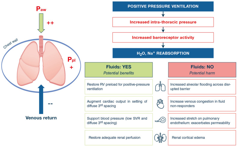
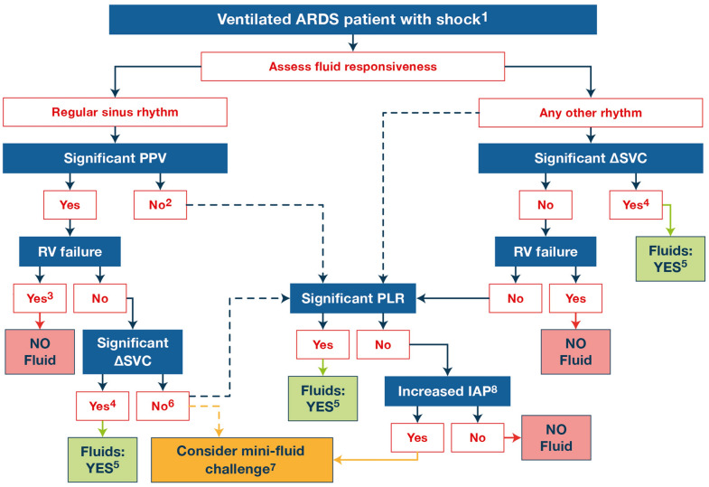
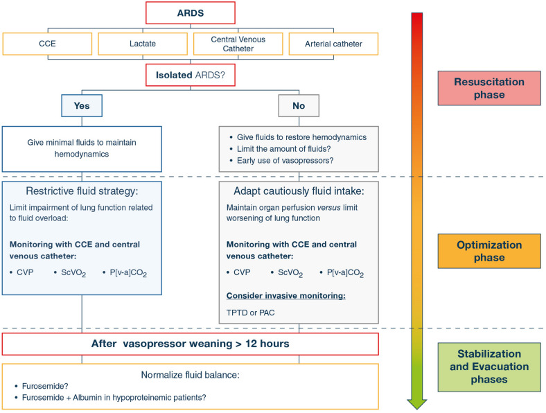
Modalities of fluid management in patients sustaining the acute respiratory distress syndrome (ARDS) are challenging and controversial. Optimal fluid management should provide adequate oxygen delivery to the body, while avoiding inadvertent increase in lung edema which further impairs gas exchange. In ARDS patients, positive fluid balance has been associated with prolonged mechanical ventilation, longer ICU and hospital stay, and higher mortality. Accordingly, a restrictive strategy has been compared to a more liberal approach in randomized controlled trials conducted in various clinical settings. Restrictive strategies included fluid restriction guided by the monitoring of extravascular lung water, pulmonary capillary wedge or central venous pressure, and furosemide targeted to diuresis and/or albumin replacement in hypoproteinemic patients. Overall, restrictive strategies improved oxygenation significantly and reduced duration of mechanical ventilation, but had no significant effect on mortality. Fluid management may require different approaches depending on the time course of ARDS (i.e., early vs. late period). The effects of fluid strategy management according to ARDS phenotypes remain to be evaluated. Since ARDS is frequently associated with sepsis-induced acute circulatory failure, the prediction of fluid responsiveness is crucial in these patients to avoid hemodynamically inefficient-hence respiratory detrimental-fluid administration. Specific hemodynamic indices of fluid responsiveness or mini-fluid challenges should be preferably used. Since the positive airway pressure contributes to positive fluid balance in ventilated ARDS patients, it should be kept as low as possible. As soon as the hemodynamic status is stabilized, correction of cumulated fluid retention may rely on diuretics administration or renal replacement therapy.

Keywords: Acute respiratory distress syndrome; Fluid therapies; Prognosis; Pulmonary edema; Water–electrolyte balance.


Conflict of interest statement

PV, BE, PA, MB, SC, JD, GG, MJ, GSM, LG and DC: no conflict of interest. CSC: research funding from the National Institutes of Health and Roche/Genentech (present), Bayer (past); consultancies for Quark, Vasomune, and Gen1e Life Sciences.

Figures

Fig. 1
Fig. 1
Schematic representation of summarized effects of positive-pressure ventilation on fluid balance and of the potential benefit–risk ratio of fluid administration in patients with the acute respiratory distress syndrome. H2O water, Na+ sodium, Paw airway pressure, Ppl pleural pressure
Fig. 2
Fig. 2
Summary of hemodynamic parameters available to predict fluid responsiveness in ventilated patients with the acute respiratory distress syndrome. The color code reflects the advantages (green) and drawbacks (red) of each test, with the orange color for neutrality. ARDS acute respiratory distress syndrome, CCE critical care echocardiography, CO cardiac output, IVC inferior vena cava, RV right ventricle, SVC superior vena cava, TEE transesophageal echocardiography, TPTD transpulmonary thermodilution. *End-expiratory occlusion with TPTD or combined end-expiratory and end-inspiratory occlusions with CCE
Fig. 3
Fig. 3
Proposed diagnostic algorithm in ventilated ARDS patients presenting with shock based on a hemodynamic assessment using critical care echocardiography during the resuscitation and optimization periods [24]. Fluid responsiveness should be assessed to avoid hemodynamically inefficient and potentially respiratory detrimental fluids administration. In patients with sinus rhythm, pulse pressure variation can be used as a warning sign for identifying the mechanism of left ventricular load dependency revealed by heart–lung interactions (i.e., right ventricular failure vs hypovolemia responsible for Δ-down; rarely severe left ventricular failure responsible for Δ-up). In patients with other cardiac rhythms, respiratory variations of the superior vena cava diameter are the most specific parameter to predict fluid responsiveness which requires transesophageal echocardiography. Alternatively, a passive leg raising may be used to assess fluid responsiveness. When values of hemodynamic indices are within the “grey zone” or in the presence of increased intra-abdominal pressure (risk of false-negative result), mini-fluid challenges may be considered. 1The diagnostic workup must include the precise clinical setting, ongoing therapy, clinical hemodynamic assessment and biological markers of tissue hypoperfusion. In ARDS patients, specific parameters of fluid responsiveness should be preferred.2When pulse pressure variation is in the “grey zone”, a passive leg raising may be performed to seek for fluid responsiveness. 3Right ventricular failure typically associates an acute dilatation of the right ventricular cavity and increased central venous pressure secondary to systemic venous congestion [36]. 4In ARDS patients, a ΔSVC cut-off of 31% predicts fluid responsiveness with a 90% specificity, at the expense of a low sensitivity of 43% [42]. Associated echocardiography findings consistent with decreased cardiac preload are frequently associated (e.g., hyperkinetic right ventricle with small cavity size, decreased diameter of the inferior vena cava with marked respiratory variation, significant respiratory variation of maximal left ventricular outflow tract Doppler velocity [41]. 5Repeated small aliquots (e.g., 250 mL) are preferable; both efficacy (percentage of increase of left ventricular stroke volume when compared to baseline) and tolerance (absence of significant increase in left ventricular filling pressure) of fluid challenge should be assessed using serial hemodynamic assessment. 6To increase the sensitivity of superior vena cava respiratory variation, lower threshold value can be used (e.g. a 4% cut-off has a sensitivity of 89%) [42], or a mini-fluid challenge can be considered. 7: mini-fluid challenges consist in administrating intravenously a small volume of fluids over a very short period of time (e.g., 50–100 mL within 1 min) [–48]. 8Intra-abdominal pressure should best be measured in patients at high risk of intra- abdominal hypertension [44] since elevated values may result in falsely negative passive leg raising [37]. ARDS acute respiratory distress syndrome, ΔSVC respiratory variation of the superior vena cava diameter, IAP intra-abdominal pressure, PPV pulse pressure variation, RV right ventricle
Fig. 4
Fig. 4
Proposed practical management of fluid balance according to the cause and timing of ARDS according to the four-hit theory/ROSE concept [24]. CVP central venous pressure, PAC pulmonary artery catheter, CCE critical care echocardiography, ScVO2 central venous oxygen saturation, P[v-a]CO2 veno-arterial carbon dioxide tension difference, TPTD transpulmonary thermodilution

References

    1. Vieillard-Baron A, Matthay M, Teboul JL, Bein T, Schultz M, Magder S, Marini JJ. Experts' opinion on management of hemodynamics in ARDS patients: focus on the effects of mechanical ventilation. Intensive Care Med. 2016;42:739–749. - PubMed
    1. Jia X, Malhotra A, Saeed M, Mark RG, Talmor D. Risk factors for ARDS in patients receiving mechanical ventilation for > 48 h. Chest. 2008;133:853–861. - PMC - PubMed
    1. Simmons RS, Berdine GG, Seidenfeld JJ, Prihoda TJ, Harris GD, Smith JD, Gilbert TJ, Mota E, Johanson WG., Jr Fluid balance and the adult respiratory distress syndrome. Am Rev Respir Dis. 1987;135:924–929. - PubMed
    1. Matthay MA, Zemans RL, Zimmerman GA, Arabi YM, Beitler JR, Mercat A, Herridge M, Randolph AG, Calfee CS. Acute respiratory distress syndrome. Nat Rev Dis Primers. 2019;5:18. - PMC - PubMed
    1. Mangialardi RJ, Martin GS, Bernard GR, Wheeler AP, Christman BW, Dupont WD, Higgins SB, Swindell BB. Hypoproteinemia predicts acute respiratory distress syndrome development, weight gain, and death in patients with sepsis. Ibuprofen in Sepsis Study Group. Crit Care Med. 2000;28:3137–3145. - PubMed