Review
. 2021 Mar;250(3):393-413.
doi: 10.1002/dvdy.269. Epub 2020 Nov 21.

Bringing tendon biology to heel: Leveraging mechanisms of tendon development, healing, and regeneration to advance therapeutic strategies

Affiliations
Review

Bringing tendon biology to heel: Leveraging mechanisms of tendon development, healing, and regeneration to advance therapeutic strategies

Stephanie L Tsai et al. Dev Dyn. 2021 Mar.

Abstract

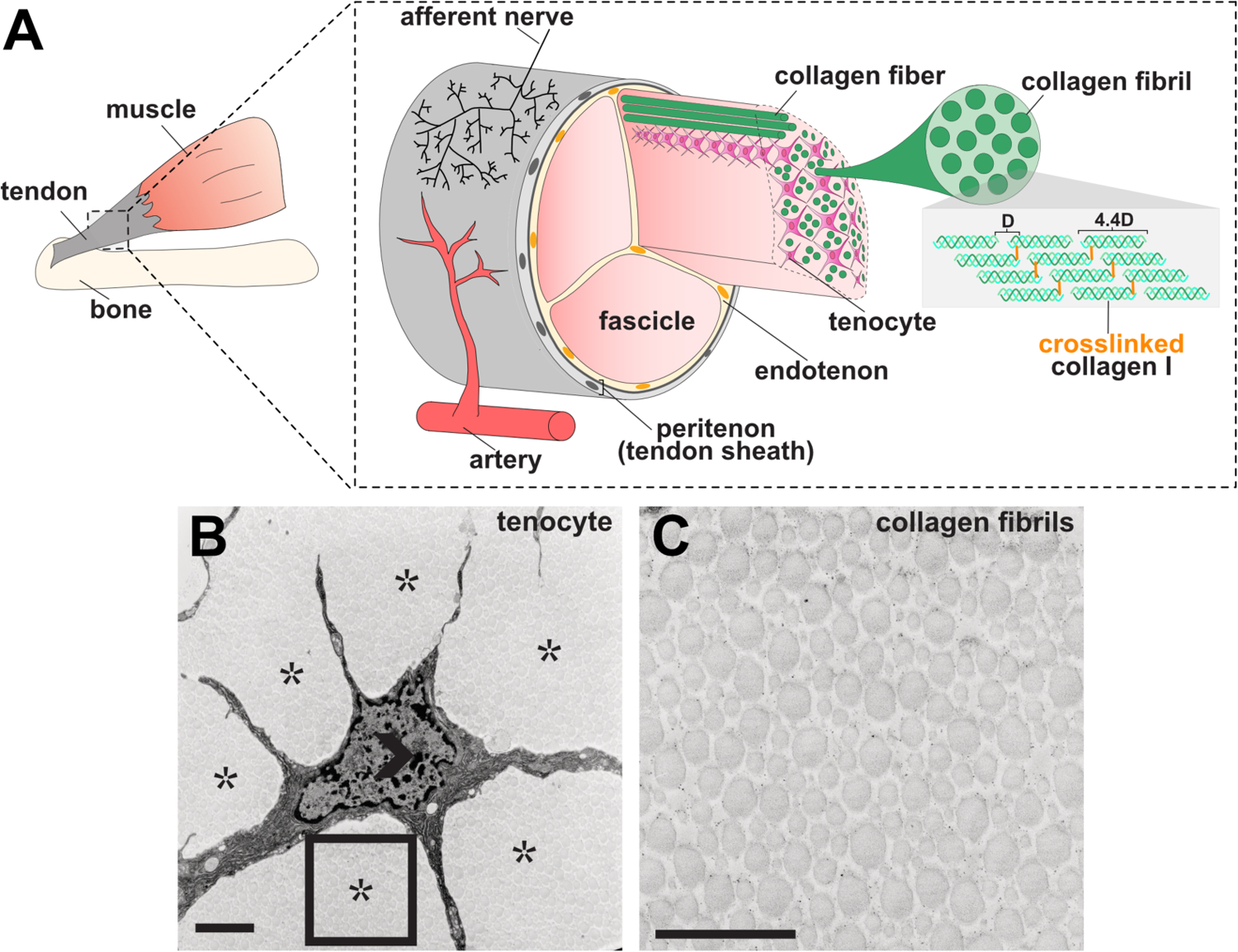
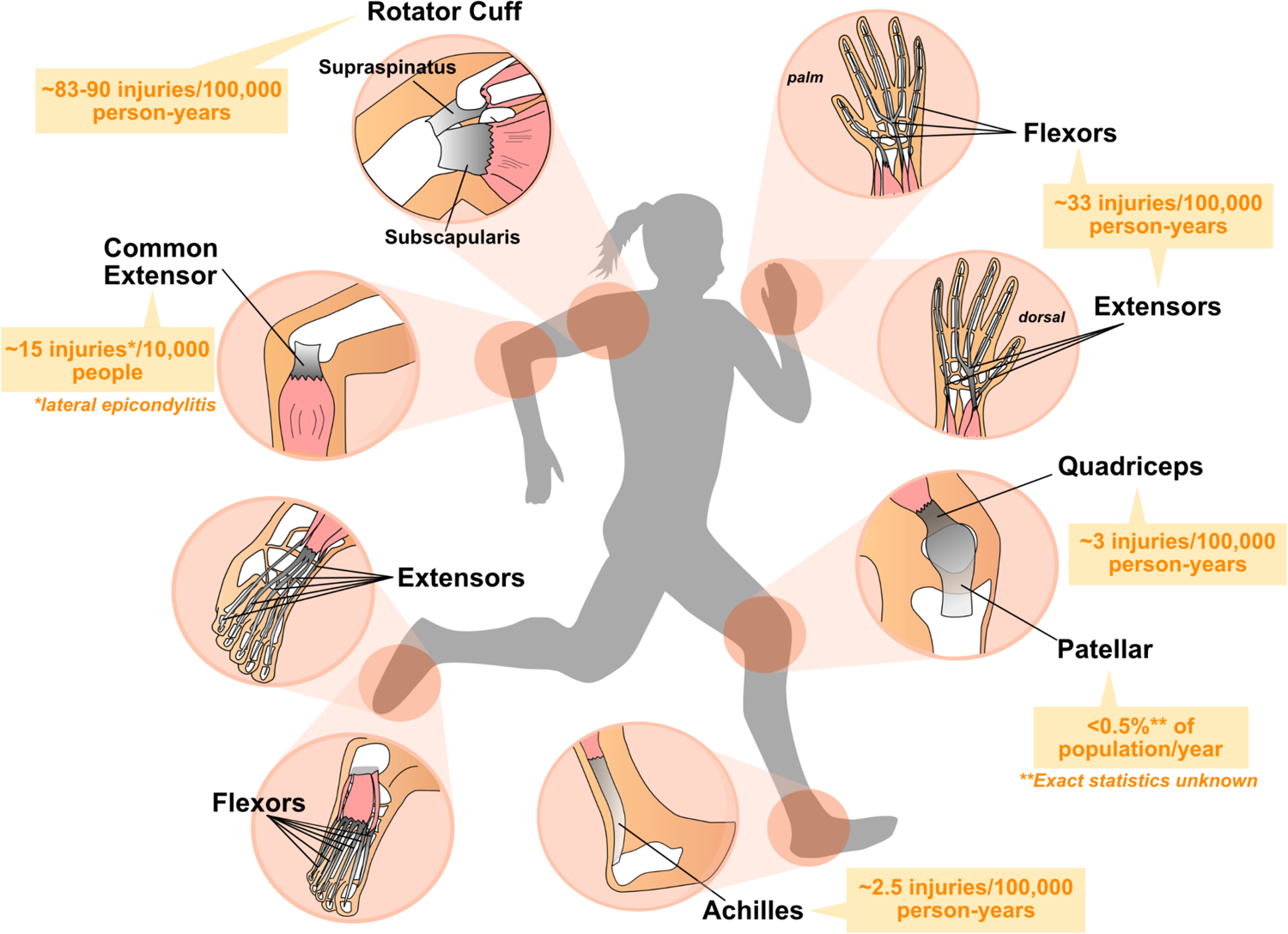
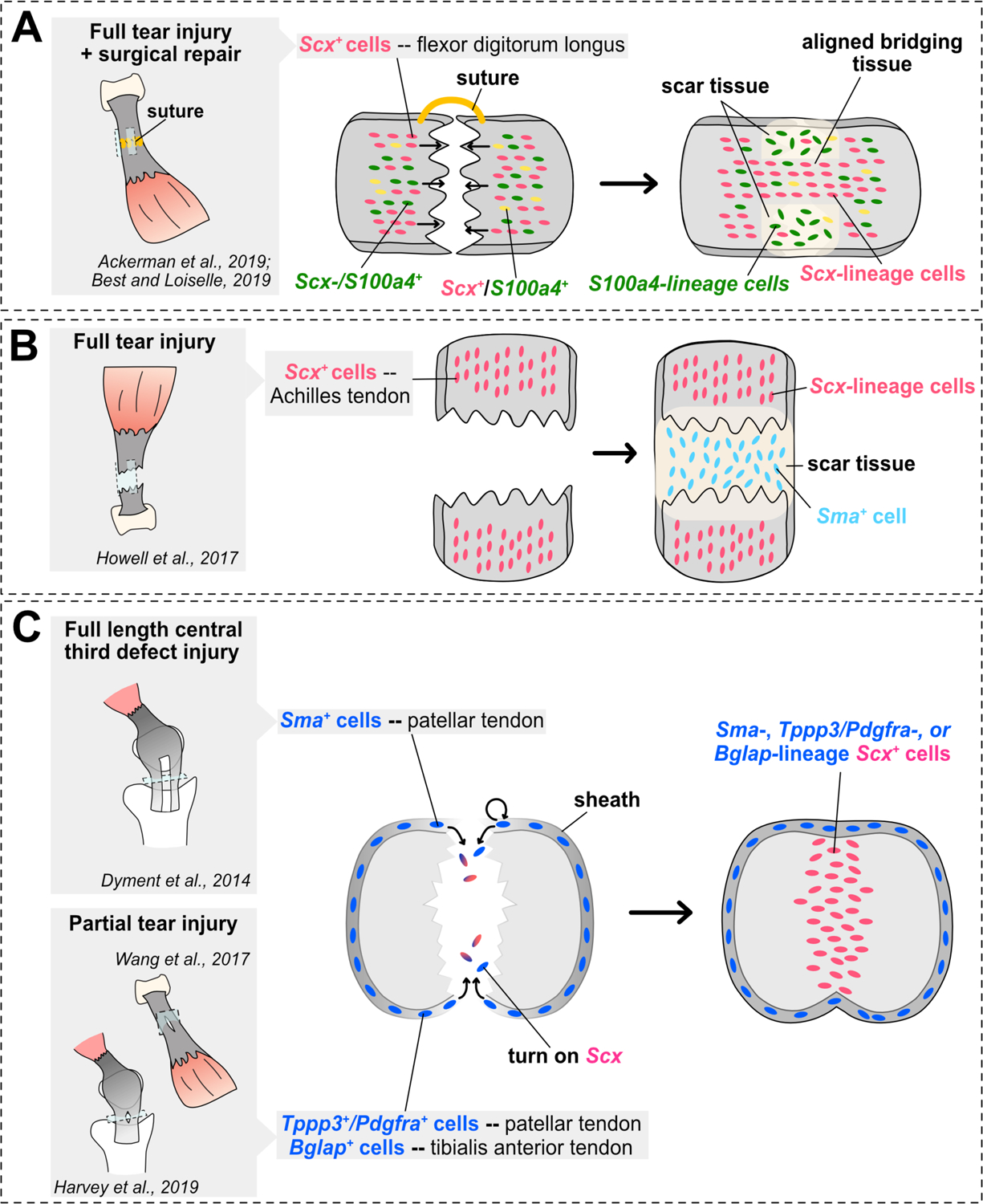
Tendons are specialized matrix-rich connective tissues that transmit forces from muscle to bone and are essential for movement. As tissues that frequently transfer large mechanical loads, tendons are commonly injured in patients of all ages. Following injury, mammalian tendons heal poorly through a slow process that forms disorganized fibrotic scar tissue with inferior biomechanical function. Current treatments are limited and patients can be left with a weaker tendon that is likely to rerupture and an increased chance of developing degenerative conditions. More effective, alternative treatments are needed. However, our current understanding of tendon biology remains limited. Here, we emphasize why expanding our knowledge of tendon development, healing, and regeneration is imperative for advancing tendon regenerative medicine. We provide a comprehensive review of the current mechanisms governing tendon development and healing and further highlight recent work in regenerative tendon models including the neonatal mouse and zebrafish. Importantly, we discuss how present and future discoveries can be applied to both augment current treatments and design novel strategies to treat tendon injuries.

Keywords: connective tissues; injury; repair; stem cells; tendon.


Figures

Figure 1.
Figure 1.. Tendon structure.
(A) Graphical representation of tendon morphology. The tendon midsubstance is comprised of collagen molecules which are spaced apart at a distance of 67 nm (letter D in diagram) and cross-linked to form stable fibrils. Tenocytes are interspersed between collagen fibrils, which together generally form higher order bundles called fascicles. Fascicles are held together by connective tissue called the endotenon. The tendon midsubstance is encased in the peritenon, or tendon sheath, which is comprised of a basement membrane and epithelial cell layer. (B–C) Transmission electron micrograph (TEM) of 6-week old mouse tenocyte (B) and collagen fibrils (C). Open arrowhead marks cell nuclei and asterisks mark collagen fibrils. Rectangle in B is shown at higher magnification in C. Scale bar, 1 um. Images in B–C were adapted from Kalson et al. (2015).
Figure 2.
Figure 2.. Major force-bearing limb tendons in the human body.
Diagram depicting the structure of major limb tendons that are frequently injured including the digital flexor and extensor tendons, common extensor, Achilles, rotator cuff, and patellar tendons. Muscles are depicted in pink, tendons in grey, and bone in white. Average incidence of injuries amongst the general population (predominantly US statistics) are shown in orange. Note: Rates of tendon rupture or disease vary with age and population demographics of geographical location or study.
Figure 3.
Figure 3.. Summary of lineage tracing studies in mouse tendon healing models.
(A–C) Schematics illustrating main conclusions of mouse tendon healing genetic lineage tracing studies in various tendon midsubstance injury models and limb tendons. Studies assessing contribution of cells from the midsubstance are shown in A–B, while studies assessing contribution of cells from the tendon sheath are shown in C. Sagittal views are shown in the cartoons in A–B, while a transverse view is shown in C.

References

    1. Komi PV. Relevance of in vivo force measurements to human biomechanics. J Biomech. 1990;23Suppl 1:23–34. 10.1016/0021-9290(90)90038-5. - DOI - PubMed
    1. Komi PV, Fukashiro S, Jarvinen M. Biomechanical loading of Achilles tendon during normal locomotion. Clin Sports Med. July1992;11(3):521–31. - PubMed
    1. Grinstein M, Dingwall HL, O’Connor LD, Zou K, Capellini TD, Galloway JL. A distinct transition from cell growth to physiological homeostasis in the tendon. Elife. September192019;8. 10.7554/eLife.48689. - DOI - PMC - PubMed
    1. Butler DL, Juncosa N, Dressler MR. Functional efficacy of tendon repair processes. Annu Rev Biomed Eng. 2004;6:303–29. 10.1146/annurev.bioeng.6.040803.140240. - DOI - PubMed
    1. Nourissat G, Berenbaum F, Duprez D. Tendon injury: from biology to tendon repair. Nat Rev Rheumatol. April2015;11(4):223–33. 10.1038/nrrheum.2015.26. - DOI - PubMed

Publication types