Review
. 2021 Mar;22(3):454-463.
doi: 10.3348/kjr.2020.0191. Epub 2020 Oct 30.

Interstitial Lung Abnormalities: What Radiologists Should Know

Affiliations
Review

Interstitial Lung Abnormalities: What Radiologists Should Know

Kum Ju Chae et al. Korean J Radiol. 2021 Mar.

Abstract

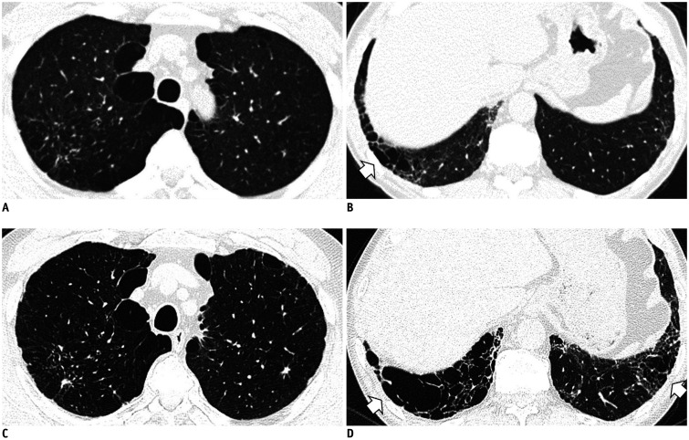
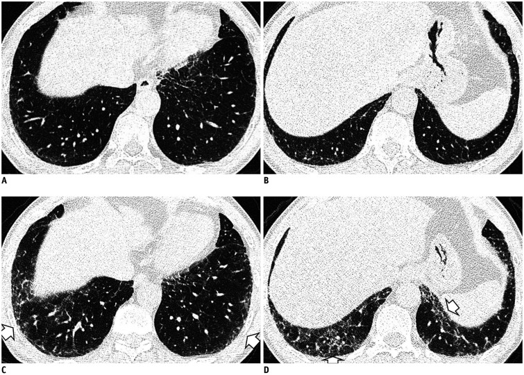
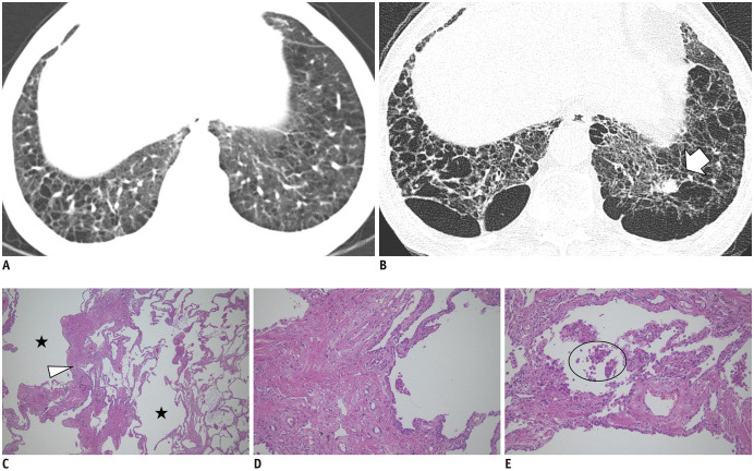
Interstitial lung abnormalities (ILAs) are radiologic abnormalities found incidentally on chest CT that are potentially related to interstitial lung diseases. Several articles have reported that ILAs are associated with increased mortality, and they can show radiologic progression. With the increased recognition of ILAs on CT, the role of radiologists in reporting them is critical. This review aims to discuss the clinical significance and radiologic characteristics of ILAs to facilitate and enhance their management.

Keywords: Idiopathic pulmonary fibrosis; Interstitial lung abnormalities; Interstitial lung disease; Smoking.


Conflict of interest statement

The authors have no potential conflicts of interest to disclose.

Figures

Fig. 1
Fig. 1. Typical image patterns demonstrating ILA.
A. A representative axial CT image showing bilateral subpleural GGA (arrows). To confirm a dependent opacity, this CT was taken in a prone position. B. CT image taken in the prone position shows bilateral subpleural reticulation in both lungs (arrow). C. Subpleural non-emphysematous cysts in both lungs. These present as thin-walled variably sized cysts on CT images (arrows). D. CT images demonstrate honeycombing in both lungs (arrows). These are clustered cystic air spaces, and their sizes are usually uniform (3–10 mm). E. Image shows traction bronchiectasis/bronchiolectasis (arrow). This can be seen as thick-walled bronchial or bronchiolar dilatation in the subpleural area of the lower lobe. GGA = ground-glass attenuation, ILA = interstitial lung abnormality
Fig. 2
Fig. 2. Dependent opacity of the lung.
A. CT image obtained in the supine position shows diffuse GGA in the subpleural area of the lower lobes and posterior basal segment (arrows). B. The GGA disappeared in the CT obtained in the prone position (arrows).
Fig. 3
Fig. 3. Fibrosis adjacent to spinal osteophytes.
A. Localized GGA and reticular opacity are seen in the right lower lobe's medial subpleura (arrow). B. Coronal reconstruction shows that the linear fibrotic change is elongated adjacent to the thoracic spine (arrow).
Fig. 4
Fig. 4. Unilateral abnormality of the lung.
CT image illustrating unilateral GGA and traction bronchiectasis of the right lower lobe (arrow). In the case of unilateral pulmonary fibrosis, it is better to consider other causes such as chronic inflammatory disease, radiation-induced injury, or circulatory disorders.
Fig. 5
Fig. 5. Diffuse centrilobular nodularity.
CT image of a current smoker (30 pack-years) shows ill-defined diffuse centrilobular nodules in the upper lobes (arrows).
Fig. 6
Fig. 6. ILA associated with connective tissue disease.
CT image of a 71-year-old female patient shows bilateral subpleural GGA with juxta subpleural sparing (arrows). After the evaluation of connective tissue disease, she was diagnosed as having systemic sclerosis-induced lung disease.
Fig. 7
Fig. 7. Occupation-induced lung fibrosis.
A. High-resolution CT image shows bilateral subpleural reticulation, honeycombing, and traction bronchiectasis (arrows). B. The mediastinal window setting image demonstrates bilateral multiple noncalcified pleural plaques (arrowheads). The patient, having a history of long-standing exposure to asbestos, was diagnosed as having asbestosis and asbestos-related pleural plaques.
Fig. 8
Fig. 8. Serial chest CT images show a progression of fibrotic ILA to UIP pattern in a 61-year-old male former smoker with 45 pack-years of cigarette consumption.
A. An initial image at the level of the posterior basal segment shows mild reticular opacity and bronchiolectasis in the subpleural zone (arrow). At this time, he had no respiratory symptoms, and CT was performed for the evaluation of an incidentally detected lung nodule on chest X-ray. B. A follow-up CT image after four years shows the progression of reticular opacity and newly developed honeycombing in the peripheral portion of the lower lung (arrows). C. After 9 years from the baseline CT, a progression of traction bronchiectasis and honeycombing of the lower lobe, which is a UIP pattern, is evident (arrows). UIP = usual interstitial pneumonia
Fig. 9
Fig. 9. Serial chest CT images show a progression of nonemphysematous cysts in a 59-year-old male former smoker with 50 pack-years of cigarette consumption.
A, B. Initial CT images show emphysema in the upper lobes and nonemphysematous cysts (arrow) in the lower lobes. C, D. After 10 years of follow-up, CTs show an increase in the extent and size of nonemphysematous cysts in the lower lobes (arrows).
Fig. 10
Fig. 10. Serial chest CT images show a progression of nonfibrotic ILA in a 57-year-old male former smoker with ten pack-years of cigarette consumption.
A, B. Initial CT images show mild reticular opacity in the subpleural zone. At this time, his lung function was within a normal range. C, D. Follow-up CT images after three years show the progression of reticular opacity and GGA in the peripheral portion of the lower lung (arrows). During this period, the patient experienced a decrease in total lung capacity (79% of predicted) and diffusion capacity (85% of predicted).
Fig. 11
Fig. 11. Chest CT images and histopathologic findings of nonemphysematous cysts in a 60-year-old current smoker.
A. The initial image shows emphysema and nonemphysematous cysts in the lower lobes. B. After 12 years, the CT image shows the progression of emphysema and nonemphysematous cysts, with a newly developed lung nodule in the left lower lobe (arrow), which was proven to be a squamous cell carcinoma by left lower lobectomy. C. Low magnification (× 40) of the histopathologic finding shows marked interstitial thickening (arrowhead) with prominent emphysema (asterisks). D, E. High magnification (× 100) findings show interstitial thickening consists of thick collagen bundles mixed with hyperplastic smooth muscle fibers and pigmented macrophages (circle) are present in air spaces. The histopathologic findings are consistent with smoking-related interstitial fibrosis, which is a pathologic subtype of ILAs (hematoxylin & eosin staining).

References

    1. Hatabu H, Hunninghake GM, Lynch DA. Interstitial lung abnormality: recognition and perspectives. Radiology. 2019;291:1–3. - PubMed
    1. Hobbs BD, Putman RK, Araki T, Nishino M, Gudmundsson G, Gudnason V, et al. Overlap of genetic risk between interstitial lung abnormalities and idiopathic pulmonary fibrosis. Am J Respir Crit Care Med. 2019;200:1402–1413. - PMC - PubMed
    1. Wells AU, Kokosi MA. Subclinical interstitial lung abnormalities: toward the early detection of idiopathic pulmonary fibrosis? Am J Respir Crit Care Med. 2016;194:1445–1446. - PMC - PubMed
    1. Seibold MA, Wise AL, Speer MC, Steele MP, Brown KK, Loyd JE, et al. A common MUC5B promoter polymorphism and pulmonary fibrosis. N Engl J Med. 2011;364:1503–1512. - PMC - PubMed
    1. Doyle TJ, Hunninghake GM, Rosas IO. Subclinical interstitial lung disease: why you should care. Am J Respir Crit Care Med. 2012;185:1147–1153. - PMC - PubMed

MeSH terms