Review
. 2020 Nov 17;9(22):e018275.
doi: 10.1161/JAHA.120.018275. Epub 2020 Nov 10.

Systematic Approach to Malalignment Type Ventricular Septal Defects

Affiliations
Review

Systematic Approach to Malalignment Type Ventricular Septal Defects

Shi-Joon Yoo et al. J Am Heart Assoc. .

Abstract

Various congenital heart diseases are associated with malalignment of a part of the ventricular septum. Most commonly, the outlet septum is malaligned toward the right or left ventricle. Less commonly, the whole or a major part of the ventricular septum is malaligned in relation to the atrial septal plane. Although the pathological conditions associated with ventricular septal malalignment have been well recognized, the descriptions are often confusing and sometimes incorrect. In this pictorial essay, we introduce our systematic approach to the assessment of malalignment type ventricular septal defects with typical case examples. The systematic approach comprises description of the essential features of malalignment, including the following: (1) the malaligned part of the ventricular septum, (2) the reference structure, (3) the mechanism of malalignment, (4) the direction of malalignment, and (5) the severity of malalignment.

Keywords: double outlet right ventricle; malalignment; overriding; straddling; ventricular septal defect.


Conflict of interest statement

None.

Figures
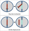
Figure 1
Figure 1. Flap‐door vs en‐bloc displacement mechanisms of ventricular septal malalignment.
In flap‐door mechanism, the malaligned part of the septum remains inserted to a margin of the ventricular septal defect. In en‐bloc displacement mechanism, the malaligned part of the septum is parallel with the rest of the septum.
Figure 2
Figure 2. Twisting.
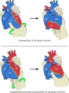
The cardiac chambers and arterial trunks show unusual spatial relationship as if the heart is twisted around the long axis of the heart. The hearts with D‐loop ventricles typically appear twisted clockwise by the observer’s right hand on the apex while the back of the heart is held by the observer’s left hand. The hearts with L‐loop ventricles typically appear twisted counterclockwise. The atrial and ventricular septa are twisted and malaligned, with one or both atrioventricular valves frequently overriding/straddling the ventricular septum. Data derived from Yoo, et al. 13 Ao indicates aorta; LA, left atrium; LV, left ventricle; PT, pulmonary arterial trunk; RA, right atrium; and RV, right ventricle.
Figure 3
Figure 3. Cartoons showing the flap‐door mechanism of malalignment of the outlet septum in tetralogy of Fallot.
The door of the outlet septum opens into the right ventricle (RV) with the hinges on the left side, resulting in subpulmonary outflow tract narrowing, small size of the pulmonary valve, and overriding of the large aortic valve (AV). Ao indicates aorta; LV, left ventricle; MV, mitral valve; PT, pulmonary arterial trunk; RA, right atrium; RV, right ventricle; TSM, trabecula septomarginalis; and TV, tricuspid valve.
Figure 4
Figure 4. Cartoons showing the flap‐door mechanism of malalignment of the outlet septum in so‐called interruption or coarctation type of ventricular septal defect.
The door of the outlet septum opens into the left ventricle (LV) (red arrow) with the hinges on the top along the ventriculoinfundibular fold/aortic valve attachment, resulting in narrowing of the subaortic outflow tract and small aortic valve. Ao indicates aorta; LA, left atrium; PDA, patent ductus arteriosus; PT, pulmonary arterial trunk; RA, right atrium; RV, right ventricle; TSM, trabecula septomarginalis; and TV, tricuspid valve.
Figure 5
Figure 5. Short axis cuts of the ventricles seen from the apex, showing the spectrum of tricuspid valve (TV) overriding/straddling and double inlet left ventricle (LV) in the setting of situs solitus, D‐loop ventricles, and normally related and connected great arteries.
The ventricular septum is malaligned like a flap door, with the hinges at the anterior interventricular groove and the posterior insertion offset from the crux cordis (CC) posteriorly. The green dotted arrows indicate the direction and severity of malalignment. The ventricular septal defect is marked with red dotted lines. An elevated muscular ridge, called the posteromedian muscle ridge (PMM), is usually seen along the posterior wall of the LV inlet between the 2 atrioventricular valves. The classic positions of the papillary muscles are marked with circles in the lower panel diagrams. Ant indicates anterior; Lt, left; MV, mitral valve; Post, posterior; Rt, right; and RV, right ventricle.
Figure 6
Figure 6. Short axis cuts of the ventricles seen from the apex, showing the spectrum of tricuspid valve (TV) overriding/straddling and double inlet left ventricle (LV) in the setting of situs solitus, L‐loop ventricles, and transposed/malposed great arteries.
The ventricular septum is malaligned like a flap door with the hinges at the anterior interventricular groove and the posterior insertion offset from the crux cordis (CC) posteriorly. The green dotted arrows indicate the direction and severity of malalignment. The ventricular septal defect is marked with red dotted lines. An elevated muscular ridge, called the posteromedian muscle ridge (PMM), is usually seen along the posterior wall of the LV inlet between the 2 atrioventricular valves. The classic positions of the papillary muscles are marked with circles in the lower panel diagrams. Ant indicates anterior; Lt, left; MV, mitral valve; Post, posterior; Rt, right; and RV, right ventricle.
Figure 7
Figure 7. Short axis cuts of the ventricles seen from the apex, showing the spectrum of mitral valve (MV) overriding/straddling and double inlet right ventricle (RV) in the setting of situs solitus, D‐loop ventricles, and transposed/malposed great arteries.
The ventricular septum is malaligned like a flap door with the hinges at the crux cordis (CC) and posterior interventricular groove and the anterior interventricular groove deviated toward the side of the left ventricle (LV). The green dotted arrows indicate the direction and severity of malalignment. The ventricular septal defect is marked with red dotted lines. The classic positions of the papillary muscles are marked with circles in the lower panel diagrams. Ant indicates anterior; Lt, left; Post, posterior; Rt, right; and TV, tricuspid valve.

References

    1. Anderson RH, Wilcox BR. The surgical anatomy of ventricular septal defects associated with overriding valvar orifices. J Card Surg. 1993;8:130–142. - PubMed
    1. Spicer DE, Hsu HH, Co‐Vu J, Anderson RH, Ficker FJ. Ventricular septal defect. Orphanet J Rare Dis. 2014;9:144–159. - PMC - PubMed
    1. Crucean A, Brawn WJ, Spicer DE, Franklin RC, Anderson RH. Holes and channels between the ventricles revisited. Cardiol Young. 2016;25:1099–1110. - PubMed
    1. Bharati S, McAllister HA Jr, Lev M. Straddling and displaced atrioventricular orifices and valves. Circulation. 1979;60:673–684. - PubMed
    1. Milo S, Ho SY, Macartney FJ, Wilkinson JL, Becker AE, Wenink AC, Gittenberger de Groot AC, Anderson RH. Straddling and overriding atrioventricular valves: morphology and classification. Am J Cardiol. 1979;44:1122–1134. - PubMed

MeSH terms

LinkOut - more resources