. 2021 Feb;191(2):274-282.
doi: 10.1016/j.ajpath.2020.10.016. Epub 2020 Nov 7.

Acute Respiratory Distress in Aged, SARS-CoV-2-Infected African Green Monkeys but Not Rhesus Macaques

Affiliations

Acute Respiratory Distress in Aged, SARS-CoV-2-Infected African Green Monkeys but Not Rhesus Macaques

Robert V Blair et al. Am J Pathol. 2021 Feb.

Abstract

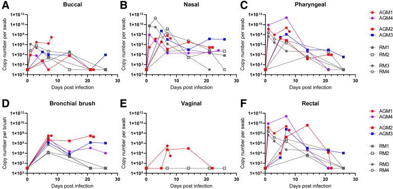
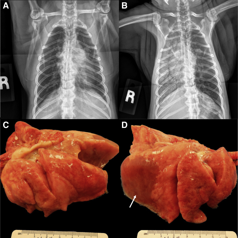
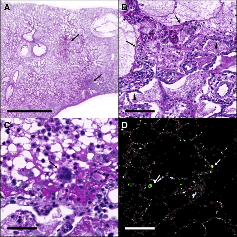
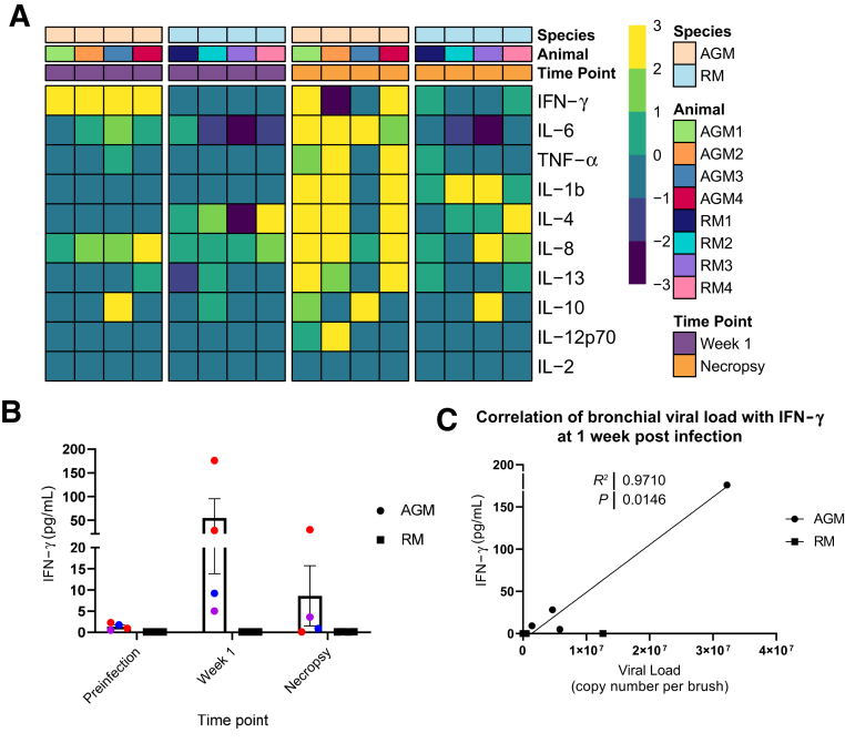
Severe acute respiratory syndrome coronavirus 2 (SARS-CoV-2) induces a wide range of disease severity, ranging from asymptomatic infection to a life-threating illness, particularly in the elderly population and individuals with comorbid conditions. Among individuals with serious coronavirus 2019 (COVID-19) disease, acute respiratory distress syndrome (ARDS) is a common and often fatal presentation. Animal models of SARS-CoV-2 infection that manifest severe disease are needed to investigate the pathogenesis of COVID-19-induced ARDS and evaluate therapeutic strategies. We report two cases of ARDS in two aged African green monkeys (AGMs) infected with SARS-CoV-2 that had pathological lesions and disease similar to severe COVID-19 in humans. We also report a comparatively mild COVID-19 phenotype characterized by minor clinical, radiographic, and histopathologic changes in the two surviving, aged AGMs and four rhesus macaques (RMs) infected with SARS-CoV-2. Notable increases in circulating cytokines were observed in three of four infected, aged AGMs but not in infected RMs. All the AGMs had increased levels of plasma IL-6 compared with baseline, a predictive marker and presumptive therapeutic target in humans infected with SARS-CoV-2. Together, our results indicate that both RMs and AGMs are capable of modeling SARS-CoV-2 infection and suggest that aged AGMs may be useful for modeling severe disease manifestations, including ARDS.


Figures

Figure 1
Figure 1
Quantification of viral loads from mucosal swabs. A–F; All animals [four African green monkeys (AGMs) and four rhesus macaques (RMs)] had detectable viral RNA at mucosal sites, including buccal (A), nasal (B), pharyngeal (C), bronchial brush (D), and rectal swabs (F). D: No significant differences are seen in viral load between species and route of exposure (Mann-Whitney U-test). Animals with acute respiratory disease syndrome (ARDS) trend to high viral loads in bronchial brush samples. E: Viral RNA is detected in vaginal swabs from both female AGMs but not the only female RM. Circles indicate aerosol exposure; squares, multiroute exposure; gray, RMs; red, developed ARDS; purple, increased cytokines without ARDS; and blue, no cytokine increase or ARDS.
Figure 2
Figure 2
Radiographic and gross pathologic changes in SARS-CoV-2 exposed African green monkey (AGM) 1. A and B: Radiographs 22 hours before (A) and at the time of necropsy (B) showing the rapid development of alveolar lung opacities throughout the right lung lobes. C: The left lung lobes fail to collapse. D: There is extensive consolidation of the right lower lung lobe with pulmonary edema (arrow). The right middle and anterior lobes are less affected. On cut surface, all lobes ooze copious fluid.
Figure 3
Figure 3
Histopathology and fluorescent immunohistochemistry in African green monkey (AGM) 1. A: The right lower lung lobe is filled with fibrin and edema with areas of hemorrhage and necrosis (arrows). B: Alveoli are variably lined by hyaline membranes (arrows) and type II pneumocytes (arrowheads). C: Rare multinucleated syncytia (arrow) are scattered throughout the affected lungs; D: Fluorescent immunohistochemistry for SARS-CoV-2 nucleoprotein (green, arrows) and ACE2 (red) identified low numbers of SARS-CoV-2 positive cells within the affected lung lobes. White indicates DAPI/nuclei; green, CoV-2; red, ACE2; and blue, Empty. Scale bars: 5 mm (A); 100 μm (B and D); 50 μm (C).
Figure 4
Figure 4
Cytokine increase. Heat maps (A) separated by species (first row), animal (second row), and timepoint (third row) showing changes in the levels of 10 cytokines in plasma at week 1 (purple, first two heat maps) and necropsy (orange, third and fourth heat maps) with respect to the baseline in African green monkeys (AGMs) and rhesus macaques (RMs). AGMs at 1 week post-infection (heat map 1) show increased levels of interferon (IFN)-γ, which is not observed in RMs at the same timepoint (heat map 2). At necropsy, three of the four AGMs exhibit elevated levels of multiple plasma cytokines (heat map 3) in contrast to RMs (heat map 4). Data are normalized (log2). B: Levels of IFN-γ in plasma at baseline, week 1, and at necropsy. C: Association between IFN-γ levels at week 1 and viral load in bronchial brushes (Pearson test). TNF-α, tumor necrosis factor-α.

References

    1. Wu Z., McGoogan J.M. Characteristics of and important lessons from the coronavirus disease 2019 (COVID-19) outbreak in China: summary of a report of 72314 cases from the Chinese Center for Disease Control and Prevention. JAMA. 2020;323:1239–1242. - PubMed
    1. Yang X., Yu Y., Xu J., Shu H., Xia J., Liu H., Wu Y., Zhang L., Yu Z., Fang M., Yu T., Wang Y., Pan S., Zou X., Yuan S., Shang Y. Clinical course and outcomes of critically ill patients with SARS-CoV-2 pneumonia in Wuhan, China: a single-centered, retrospective, observational study. Lancet Respir Med. 2020;8:475–481. - PMC - PubMed
    1. Chien J.Y., Hsueh P.R., Cheng W.C., Yu C.J., Yang P.C. Temporal changes in cytokine/chemokine profiles and pulmonary involvement in severe acute respiratory syndrome. Respirology. 2006;11:715–722. - PMC - PubMed
    1. Channappanavar R., Perlman S. Pathogenic human coronavirus infections: causes and consequences of cytokine storm and immunopathology. Semin Immunopathol. 2017;39:529–539. - PMC - PubMed
    1. Weiss S.R., Leibowitz J.L. Coronavirus pathogenesis. Adv Virus Res. 2011;81:85–164. - PMC - PubMed