. 2020 Nov 8;8(11):1753.
doi: 10.3390/microorganisms8111753.

Extracellular Vesicles Released by Enterovirus-Infected EndoC-βH1 Cells Mediate Non-Lytic Viral Spread

Affiliations

Extracellular Vesicles Released by Enterovirus-Infected EndoC-βH1 Cells Mediate Non-Lytic Viral Spread

Eitan Netanyah et al. Microorganisms. .

Abstract

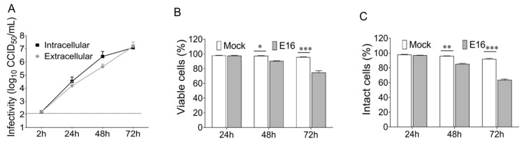
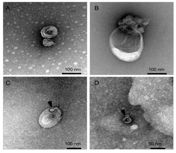
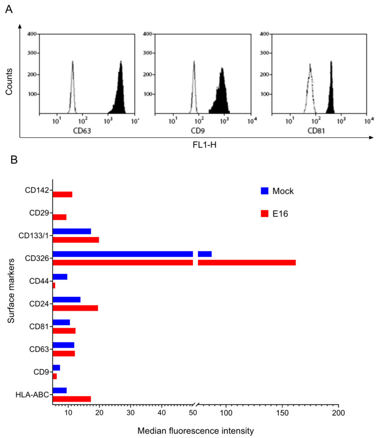
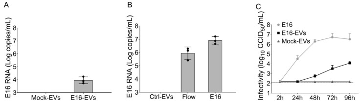
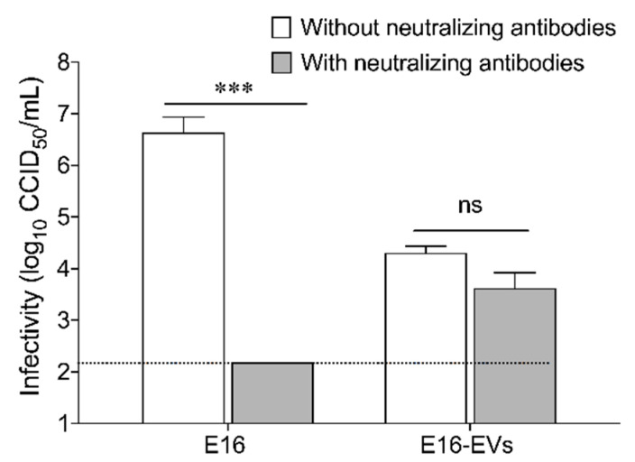
While human enteroviruses are generally regarded as a lytic virus, and persistent non-cytolytic enterovirus infection in pancreatic beta cells has been suspected of playing a role in type 1 diabetes pathogenesis. However, it is still unclear how enteroviruses could exit the pancreatic beta cell in a non-lytic manner. This study aimed to investigate the role of beta cell-derived extracellular vesicles (EVs) in the non-lytic enteroviral spread and infection. Size-exclusion chromatography and antibody-based immunoaffinity purification were used to isolate EVs from echovirus 16-infected human beta EndoC-βH1 cells. EVs were then characterized using transmission electron microscopy and Multiplex Bead-Based Flow Cytometry Assay. Virus production and release were quantified by 50% cell culture infectious dose (CCID50) assay and qRT-PCR. Our results showed that EVs from echovirus 16-infected EndoC-βH1 cells harbor infectious viruses and promote their spread during the pre-lytic phase of infection. Furthermore, the EVs-mediated infection was not inhibited by virus-specific neutralizing antibodies. In summary, this study demonstrated that enteroviruses could exit beta cells non-lytically within infectious EVs, thereby thwarting the access of neutralizing antibodies to viral particles. These data suggest that enterovirus transmission through EVs may contribute to viral dissemination and immune evasion in persistently infected beta cells.

Keywords: beta cells; enterovirus; exosomes; extracellular vesicles; type 1 diabetes; virus spread.


Conflict of interest statement

The authors declare no conflict of interest. The funders had no role in the design of the study; in the collection, analyses, or interpretation of data; in the writing of the manuscript, or in the decision to publish the results.

Figures

Figure 1
Figure 1
Pre-lytic release of echovirus 16 (E16) in EndoC-βH1 cells. (A) Cells and corresponding culture supernatant were harvested at indicated time points after infection of EndoC-βH1 cells with E16 (MOI = 0.00001) and the intracellular virus production and extracellular virus release was determined by 50% cell culture infectious dose (CCID50) assay in green monkey kidney (GMK) cells. Dotted black lines indicate the limit of detection. (B) Cell viability measured by 3-(4.5-dimethylthiazol-2-yl)-2.5-diphenyltetrazolium bromide (MTT) assay and (C) plasma membrane integrity of EndoC-βH1 cells by determining lactate dehydrogenase (LDH) activity in culture supernatant at the indicated time points after E16 infection (MOI = 0.00001) compared to mock-treated cells. Data are presented as mean ± SEM for three independent experiments, with each measurement performed in triplicate. * p < 0.05, ** p < 0.01, *** p < 0.001.
Figure 2
Figure 2
Representative transmission electron microscopy image of negatively stained EVs isolated from (AC) E16-infected EndoC-βH1 cells (fraction 7-9) and (D) viral stock solution (fraction 16–18) using SEC qEV columns. Black arrowheads indicate viral particles. See Figure S1 for further details.
Figure 3
Figure 3
Phenotyping of EndoC-βH1 cell-derived EVs (A) Flow cytometer results showing the expression levels of CD63, CD9, and CD81 in EndoC-βH1 cell-derived EVs. Barren areas are isotype control and shaded areas represent positive marker expression. See Figure S2 for complete datasets of all replicates (B) Median fluorescence intensities values of selected markers detected in EVs isolated from mock- and E16-infected EndoC-βH1 cells. The figure shows representative data of at least five independent experiments using bead-based multiplex flow cytometry assay. See Figure S3 for complete datasets of all replicates.
Figure 4
Figure 4
EVs released from E16-infected EndoC-βH1 cells transmit productive enterovirus infection to recipient cells. (A) Real-time qPCR analysis of enterovirus RNA content in immunopurified EpCAM-positive EVs from mock (Mock-EVs) or E16-infected EndoC-βH1 cells (E16-EVs). (B) EVs derived from mock-infected EndoC-βH1 cells were exposed to free E16 and re-isolated by the immunomagnetic selection of EpCAM-positive EVs. Enterovirus RNA content was analyzed by qPCR in EpCAM-selected EVs (Control-EVs) and flow-through samples. Cell-free virus (E16) was used as a positive control. (C) Naïve EndoC-βH1 cells were incubated with EVs from E16-infected EndoC-βH1 cells (E16-EVs) or mock-infected cells (Mock-EVs). EndoC-βH1 cells infected with the cell-free virus (E16) at an MOI of 0.00001 were used as a positive control. At 2 hpi, cells were washed off, and cultured in fresh Dulbecco’s Modified Eagle Medium (DMEM) medium for further 96 h. Total EndoC-βH1 cell lysates (cells plus supernatants) were collected at different time points and viral titers determined by end-point dilution on GMK cells. Dotted black lines indicate the limit of detection. Data are representative of three independent experiments, with each measurement performed in triplicate (mean ± SEM).
Figure 5
Figure 5
EVs-mediated E16 infection of EndoC-βH1 cells is not inhibited by virus-specific neutralizing antibodies. Free virus (E16) and EVs from E16-infected EndoC-βH1 cells (E16-EVs) were incubated with or without anti-E16 neutralizing antibodies. Titration of viral production in the presence or absence of neutralizing antibodies was determined as described in the materials and methods. Dotted black lines indicate the limit of detection. Data are representative of three independent experiments, with each measurement performed in triplicate (mean ± SEM). *** p < 0.001.

References

    1. Pallansch M., Roos R. Enteroviruses: Polioviruses, coxsackieviruses, echoviruses and newer enteroviruses. In: Knipe D.M., Howley P.M., editors. Fields Virology. 5th ed. Lippincott Williams & Wilkins; Philadelphia, PA, USA: 2006. pp. 895–910.
    1. Yeung W.C., Rawlinson W.D., Craig M.E. Enterovirus infection and type 1 diabetes mellitus: Systematic review and meta-analysis of observational molecular studies. BMJ. 2011;342:d35. doi: 10.1136/bmj.d35. - DOI - PMC - PubMed
    1. Morgan N.G., Richardson S.J. Enteroviruses as causative agents in type 1 diabetes: Loose ends or lost cause? Trends Endocrinol. Metab. 2014;25:611–619. doi: 10.1016/j.tem.2014.08.002. - DOI - PubMed
    1. Rodriguez-Calvo T. Enterovirus infection and type 1 diabetes: Unraveling the crime scene. Clin. Exp. Immunol. 2019;195:15–24. doi: 10.1111/cei.13223. - DOI - PMC - PubMed
    1. Hsu N.Y., Ilnytska O., Belov G., Santiana M., Chen Y.H., Takvorian P.M., Pau C., van der Schaar H., Kaushik-Basu N., Balla T., et al. Viral reorganization of the secretory pathway generates distinct organelles for RNA replication. Cell. 2010;141:799–811. doi: 10.1016/j.cell.2010.03.050. - DOI - PMC - PubMed

LinkOut - more resources