. 2020 Nov 7;9(11):2434.
doi: 10.3390/cells9112434.

Nanotherapeutic Modulation of Human Neural Cells and Glioblastoma in Organoids and Monocultures

Affiliations

Nanotherapeutic Modulation of Human Neural Cells and Glioblastoma in Organoids and Monocultures

Issan Zhang et al. Cells. .

Abstract

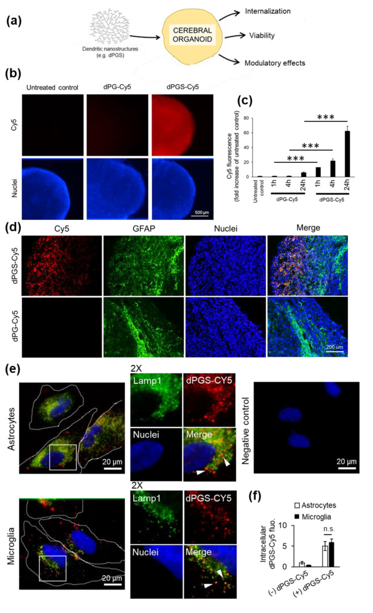
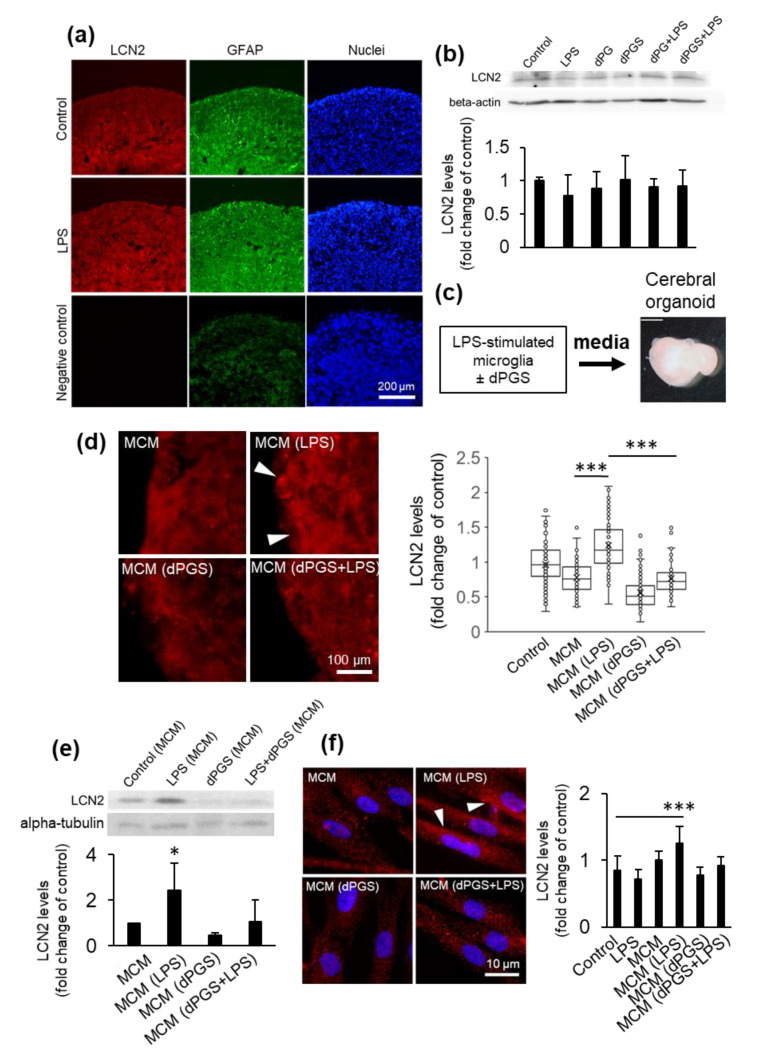
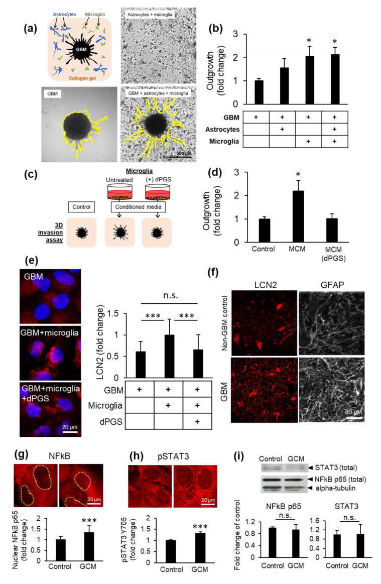
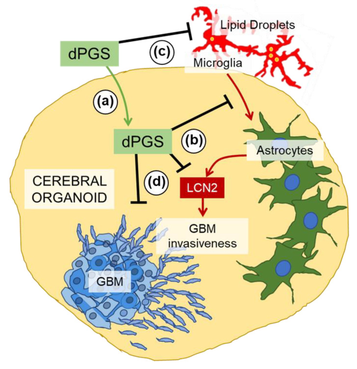
Inflammatory processes in the brain are orchestrated by microglia and astrocytes in response to activators such as pathogen-associated molecular patterns, danger-associated molecular patterns and some nanostructures. Microglia are the primary immune responders in the brain and initiate responses amplified by astrocytes through intercellular signaling. Intercellular communication between neural cells can be studied in cerebral organoids, co-cultures or in vivo. We used human cerebral organoids and glioblastoma co-cultures to study glia modulation by dendritic polyglycerol sulfate (dPGS). dPGS is an extensively studied nanostructure with inherent anti-inflammatory properties. Under inflammatory conditions, lipocalin-2 levels in astrocytes are markedly increased and indirectly enhanced by soluble factors released from hyperactive microglia. dPGS is an effective anti-inflammatory modulator of these markers. Our results show that dPGS can enter neural cells in cerebral organoids and glial cells in monocultures in a time-dependent manner. dPGS markedly reduces lipocalin-2 abundance in the neural cells. Glioblastoma tumoroids of astrocytic origin respond to activated microglia with enhanced invasiveness, whereas conditioned media from dPGS-treated microglia reduce tumoroid invasiveness. Considering that many nanostructures have only been tested in cancer cells and rodent models, experiments in human 3D cerebral organoids and co-cultures are complementary in vitro models to evaluate nanotherapeutics in the pre-clinical setting. Thoroughly characterized organoids and standardized procedures for their preparation are prerequisites to gain information of translational value in nanomedicine. This study provides data for a well-characterized dendrimer (dPGS) that modulates the activation state of human microglia implicated in brain tumor invasiveness.

Keywords: astrocytes; cerebral organoids; glioblastoma; inflammation; lipocalin-2; microglia; nanomedicines.


Conflict of interest statement

The authors declare no conflict of interest. The funders had no role in the design of the study; in the collection, analyses or interpretation of data; in the writing of the manuscript, or in the decision to publish the results.

Figures

Figure 1
Figure 1
(a) Schematic representation of cerebral organoid usage for nanostructure screening. (b) Human cerebral organoids were treated with dendritic polyglycerol (dPG)-Cy5 (1 μM) or dendritic polyglycerol sulfate (dPGS)-Cy5 (1 μM) for 1 h, 4 h or 24 h. Nuclei were labeled with Hoechst 33342. Organoids were imaged using a fluorescence microscope. (c) Cy5 fluorescence in organoids was analyzed in ImageJ. Shown are the average fluorescence per condition ± SEM. At least 90 organoids were analyzed from three independent experiments. *** p < 0.001 (d) Fluorescence micrographs of human cerebral organoids treated with dPG-Cy5 (1 μM) or dPGS-Cy5 (1 μM) for 24 h and labeled for glial fibrillary acidic protein (GFAP). Nuclei were labeled with Hoechst 33342. (e) Primary human astrocytes and human HMC3 microglia internalization of dPGS-Cy5 (1 μM) after 24 h. dPGS-Cy5 (red) is partially co-localized with Lamp1-labeled lysosomal compartments (green). The negative control was prepared in the absence of dPGS-Cy5 and primary antibody to account for background fluorescence. Nuclei were labeled with Hoechst 33342 (blue). Cells were imaged using a fluorescence microscope. (f) Shown are the average intracellular Cy5 fluorescence per cell expressed as fold change from untreated cells ± SD. At least 90 cells were analyzed in two independent experiments. n.s. Non-significant.
Figure 2
Figure 2
Lipocalin-2 levels in human cerebral organoids. (a) Micrographs of lipocalin-2 (LCN2) detected by immunofluorescence in organoid cryosections following treatment with LPS (10 ng/mL) for 24 h. Astrocytes were labeled with GFAP and nuclei with Hoechst 33342. The negative control was prepared in the absence of primary antibodies to account for background fluorescence. Samples were imaged using a fluorescence microscope. (b) LCN2 levels in organoids treated with lipopolysaccharide (LPS) (10 ng/mL) with or without dPG (1 μM) and dPGS (1 μM) for 24 h and measured by Western blot, with beta-actin as loading control. Quantification shown are the average intracellular LCN2 levels ±SD in organoids based on immunofluorescence images shown in (a). A total of 27 samples were analyzed from three independent experiments. (c) Schematic representation of media conditioning from microglia used for cerebral organoid treatment. (d) Representative fluorescence micrographs of intracellular LCN2 levels in organoids treated with conditioned media from microglia (MCM) treated with LPS (10 ng/mL), dPGS (1 μM) for 24 h. Quantifications show the average and single-cell levels of LCN2 fluorescence in cryosections. A total of 1017 cells were analyzed from independent experiments. *** p < 0.001 (e) LCN2 levels from organoids treated as in (d) and measured by Western blot, with alpha-tubulin as loading control. Shown are the average LCN2 levels from two independent experiments. * p < 0.05 (f) Fluorescence micrographs of LCN2 abundance in primary human astrocytes treated as in (d) and measured using immunocytochemistry. Shown are the average intracellular LCN2 levels in astrocytes as fold increase of the untreated control. A total of 477 cells from two independent experiments were analyzed. *** p < 0.001.
Figure 3
Figure 3
Glioblastoma (GBM) invasiveness in 3D co-cultures. (a) Collagen gels were seeded with human primary astrocytes and human HMC3 microglia in the presence or absence of a glioblastoma tumoroid. (b) Glioblastoma invasiveness in the presence or absence of microglia and astrocytes in 3D co-cultures after 6 days. Shown are the average distance of outgrowth from the tumoroid as a fold change of the tumoroid monoculture ±SEM. A total of 32 tumoroids were tested from at least three independent experiments. * p < 0.05 (c) Schematic representation of microglial conditioned media (MCM) used to treat collagen-embedded tumoroids. (d) Glioblastoma tumoroid outgrowth in the presence of conditioned media from microglia treated or not with dPGS (1 μM) after 6 days ±SEM. A total of 29 tumoroids were tested from at least four independent experiments. * p < 0.05 (e) Representative fluorescence micrographs showing intracellular LCN2 (red) in glioblastoma cells in the presence or absence of microglia cells in direct co-culture and dPGS (1 μM) for 24 h. LCN2 was fluorescently immunolabeled and cells were imaged using a fluorescence microscope. Quantifications show the average intracellular LCN2 levels ± SD per cell. At least 500 cells were analyzed from five independent experiments. *** p < 0.001; n.s. Non-significant. (f) Fluorescence micrographs of LCN2 (red) and GFAP (white) in human brain sections from non-cancerous brain or glioblastoma tumor tissues. LCN2 and GFAP were fluorescently labeled by immunohistochemistry and imaged using a fluorescence microscope. (g,h) Activation of transcription factors NFκB and STAT3 in microglia in response to glioblastoma secreted factors. Human HMC3 microglia were treated with glioblastoma conditioned media (GCM) for 24 h, after which NFκB p65 and phosphorylated STAT3 Tyr705 were fluorescently immunolabeled (red) and cells were imaged using a fluorescence microscope. Shown are (g) the average nuclear NFκB p65 level per cell ± SEM (237 cells from three independent experiments) and (h) the average pSTAT3 Y705 level per cell ± SEM (317 cells from three independent experiments) *** p < 0.001. (i) Total NFκB p65 and STAT3 protein abundance in microglia treated as in (g,h). Protein levels were determined by measurements of immunopositive bands in Western blots. Alpha-tubulin was used as loading control. Shown are the average protein abundance of NFκB p65 and STAT3 from three independent experiments. n.s. Non-significant.
Figure 4
Figure 4
Schematic representation of the proposed modulatory effects of dPGS on microglia, astrocyte and glioblastoma crosstalk. (a) dPGS is internalized in cerebral organoids, and (b) downregulates LCN2 produced by microglia-induced astrocytes, thereby (c) reducing markers of inflammation (e.g., lipid droplets) and (d) glioblastoma invasiveness.

References

    1. Paolicelli R.C., Bergamini G., Rajendran L. Cell-to-cell Communication by Extracellular Vesicles: Focus on Microglia. Neuroscience. 2019;405:148–157. doi: 10.1016/j.neuroscience.2018.04.003. - DOI - PubMed
    1. Simon E., Obst J., Gomez-Nicola D. The Evolving Dialogue of Microglia and Neurons in Alzheimer’s Disease: Microglia as Necessary Transducers of Pathology. Neuroscience. 2019;405:24–34. doi: 10.1016/j.neuroscience.2018.01.059. - DOI - PubMed
    1. Pósfai B., Cserép C., Orsolits B., Dénes Á. New Insights into Microglia–Neuron Interactions: A Neuron’s Perspective. Neuroscience. 2019;405:103–117. doi: 10.1016/j.neuroscience.2018.04.046. - DOI - PubMed
    1. Rossi G., Manfrin A., Lutolf M.P. Progress and potential in organoid research. Nat. Rev. Genet. 2018;19:671–687. doi: 10.1038/s41576-018-0051-9. - DOI - PubMed
    1. Li M., Izpisua Belmonte J.C. Organoids—Preclinical Models of Human Disease. N. Engl. J. Med. 2019;380:569–579. doi: 10.1056/NEJMra1806175. - DOI - PubMed

Publication types

Grants and funding

LinkOut - more resources