Review
. 2020 Nov 6;21(21):8323.
doi: 10.3390/ijms21218323.

Various Aspects of Calcium Signaling in the Regulation of Apoptosis, Autophagy, Cell Proliferation, and Cancer

Affiliations
Review

Various Aspects of Calcium Signaling in the Regulation of Apoptosis, Autophagy, Cell Proliferation, and Cancer

Simone Patergnani et al. Int J Mol Sci. .

Abstract

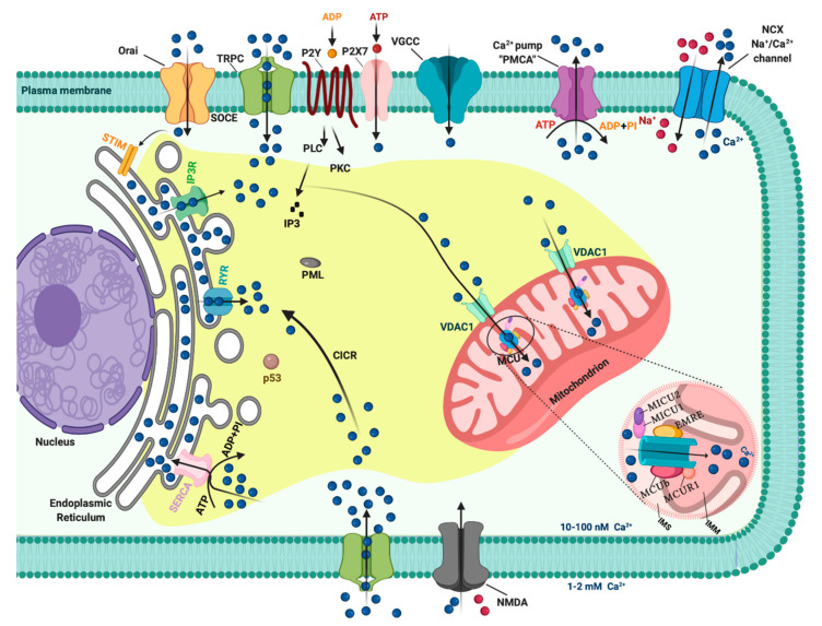
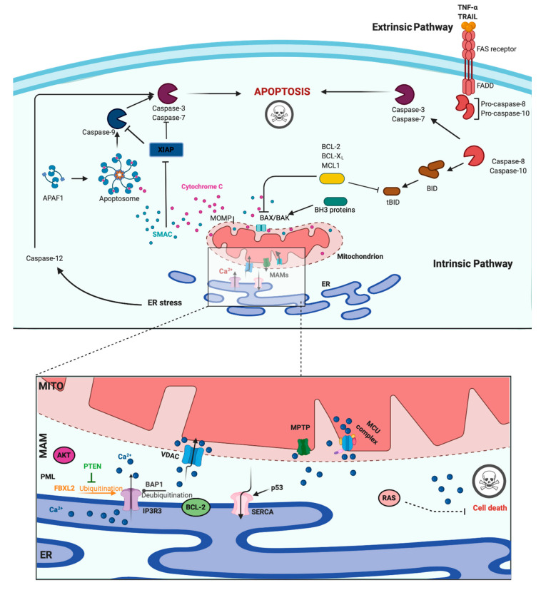
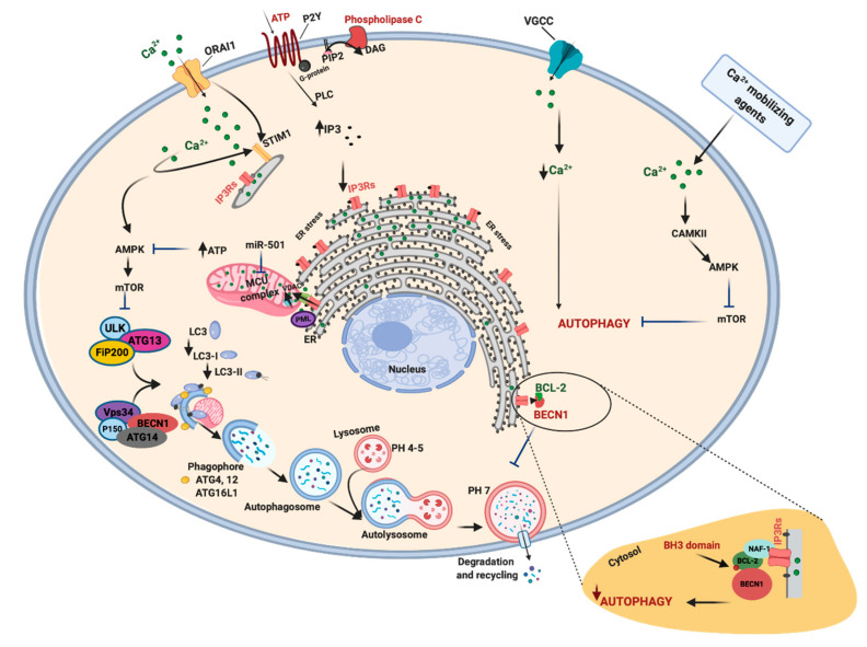
Calcium (Ca2+) is a major second messenger in cells and is essential for the fate and survival of all higher organisms. Different Ca2+ channels, pumps, or exchangers regulate variations in the duration and levels of intracellular Ca2+, which may be transient or sustained. These changes are then decoded by an elaborate toolkit of Ca2+-sensors, which translate Ca2+ signal to intracellular operational cell machinery, thereby regulating numerous Ca2+-dependent physiological processes. Alterations to Ca2+ homoeostasis and signaling are often deleterious and are associated with certain pathological states, including cancer. Altered Ca2+ transmission has been implicated in a variety of processes fundamental for the uncontrolled proliferation and invasiveness of tumor cells and other processes important for cancer progression, such as the development of resistance to cancer therapies. Here, we review what is known about Ca2+ signaling and how this fundamental second messenger regulates life and death decisions in the context of cancer, with particular attention directed to cell proliferation, apoptosis, and autophagy. We also explore the intersections of Ca2+ and the therapeutic targeting of cancer cells, summarizing the therapeutic opportunities for Ca2+ signal modulators to improve the effectiveness of current anticancer therapies.

Keywords: apoptosis; autophagy; calcium; cancer; cell cycle; chemotherapy; therapy.


Conflict of interest statement

The authors declare no conflict of interest.

Figures

Figure 1
Figure 1
The intracellular Calcium (Ca2+) signaling. Different Ca2+ transporters, channels, exchangers, binding/buffering proteins and pumps mediate the regulation of cytosolic Ca2+ concentration. In the plasma membrane (PM), PM Ca2+-ATPases (PMCA) pumps, transient receptor potential channels (TRPC), voltage-gated Ca2+ channels (VGCC), Na+/Ca2+ exchanger (NCX), and purinergic P2 receptors regulate the transport of Ca2+ ions inside and outside cells. Inositol 1,4,5-triphosphate receptors (IP3R), ryanodine receptors (RyR), and sarcoendoplasmic reticulum Ca2+-ATPase (SERCA) pumps control the storage of Ca2+ in the endoplasmic reticulum. Finally, voltage-dependent anion channels (VDAC) and members of the mitochondrial Ca2+ uniporter family are critical for controlling the mitochondrial Ca2+ uptake. Created with BioRender.com.
Figure 2
Figure 2
Apoptosis and Calcium (Ca2+) dynamics in cancer. Apoptosis is the best-characterized and studied programmed cell death. In the extrinsic pathway, extracellular ligands determine the formation of the death-inducing signaling complex that activates the caspases cascade. The intrinsic apoptotic pathway is characterized by permeabilization of the mitochondria that allows the release of cytochrome c (cyt-c) and other apoptogenic factors in the cytosol. Once released, these factors bind apoptotic protease activating factor 1 (APAF1) and form a multiprotein complex called the apoptsome that recruits and activates the caspases. Ca2+ has a major role during intrinsic apoptosis, and excessive mitochondrial Ca2+ accumulation may trigger apoptosis. Different proteins were found to control apoptotic machinery by regulating Ca2+ flux between endoplasmic reticulum (ER) and mitochondria. Antiapoptotic B-cell lymphoma-2 (BCL-2) members block apoptotic program by lowering Ca2+ levels in the ER, thereby attenuating subsequent Ca2+ release. p53 localizes at the ER–mitochondria interface to improve Ca2+ dynamics and apoptosis by increasing sarcoendoplasmic reticulum Ca2+-ATPase (SERCA) pumps activities. Additionally, the tumor suppressors promyelocytic leukemia protein (PML), BRCA1-associated protein 1 (BAP1), and phosphatase and tensin homolog (PTEN) move to the mitochondria associated membranes (MAMs) to regulate Ca2+-dependent apoptosis. They determine the activation of Ca2+ release from the ER by modulating the activity of inositol 1,4,5-triphosphate receptor 3 (IP3R3). Mutations or loss of these tumor suppressors are frequently found in diverse human tumor samples, where they lead to a reduction in Ca2+ homeostasis and the apoptosis rate, favoring cellular proliferation, tumor growth, maintenance, and metastasis. Created with BioRender.com.
Figure 3
Figure 3
Autophagy, Ca2+ and cancer. Autophagy is a key process necessary for the maintenance of the correct cell homeostasis. The unc-51 like autophagy activating kinase 1-2/autophagy-related 13/200-kDa focal adhesion kinase family-interacting protein (ULK/ATG13/FIP200) complex together with other proteins, such as coiled-coil, moesin-like BCL2 interacting protein (BECN1), controls the formation and elongation of autophagosome vesicles. The activity of BECN1 is also regulated by the portion of B-cell lymphoma-2 (BCL-2) pool that is localized in the endoplasmic reticulum (ER). A series of autophagy-related genes (ATG) is essential to the growth and closure of the autophagosome. Additionally, Ca2+ signal intervenes to modulate the autophagic machinery. A correct Ca2+ transfer between the ER and mitochondria permits optimal mitochondrial Ca2+ uptake and consequent ATP production. This signal downregulates the activation of the energy sensor of ATP/AMP ratio, 5′ adenosine monophosphate-activated protein kinase (AMPK), which is the most investigated positive regulator of autophagic induction mechanism, AMPK-ULK1-mammalian target of rapamycin (mTOR). When Ca2+ transfer from the ER to mitochondria and/or Ca2+ is imported into mitochondria is compromised, AMPK is activated, and survival autophagy is induced. For example, tumors characterized by loss of the tumor suppressor promyelocytic leukemia protein (PML) have enhanced autophagy levels and attenuated Ca2+ dynamics. In renal carcinoma, miR-501 decreases the activity of mitochondrial Ca2+ uniporter (MCU) channel, provoking a reduction in ATP production and recruitment of AMPK-ULK-mTOR pathway. In contrast, it has been observed that reduced Ca2+ dynamics may also activate autophagic cell death. Cancers have increased voltage-gated Ca2+ channels (VGCC): their inhibition reduces Ca2+ entry and activates autophagy to reduce cell proliferation. A decrease in ORAI1 delays cytoplasmic Ca2+ clearance and activates autophagy. Diverse Ca2+ mobilizing agents increase in intracellular Ca2+ levels and activate pro-survival autophagy by activating Ca2+/calmodulin-dependent protein kinases 2 (CAMKII). Created with BioRender.com.

References

    1. Raffaello A., Mammucari C., Gherardi G., Rizzuto R. Calcium at the Center of Cell Signaling: Interplay between Endoplasmic Reticulum, Mitochondria, and Lysosomes. Trends Biochem. Sci. 2016;41:1035–1049. doi: 10.1016/j.tibs.2016.09.001. - DOI - PMC - PubMed
    1. Christensen K.A., Myers J.T., Swanson J.A. pH-dependent regulation of lysosomal calcium in macrophages. J. Cell Sci. 2002;115:599–607. - PubMed
    1. Marchi S., Giorgi C., Galluzzi L., Pinton P. Ca2+ Fluxes and Cancer. Mol. Cell. 2020;78:1055–1069. doi: 10.1016/j.molcel.2020.04.017. - DOI - PubMed
    1. Bootman M.D., Bultynck G. Fundamentals of Cellular Calcium Signaling: A Primer. Cold Spring Harb. Perspect. Biol. 2020;12:a038802. doi: 10.1101/cshperspect.a038802. - DOI - PMC - PubMed
    1. Missiroli S., Perrone M., Genovese I., Pinton P., Giorgi C. Cancer metabolism and mitochondria: Finding novel mechanisms to fight tumours. EBioMedicine. 2020;59:102943. doi: 10.1016/j.ebiom.2020.102943. - DOI - PMC - PubMed

LinkOut - more resources