. 2020 Nov 6;8(11):1741.
doi: 10.3390/microorganisms8111741.

Bile Acid Signal Molecules Associate Temporally with Respiratory Inflammation and Microbiome Signatures in Clinically Stable Cystic Fibrosis Patients

Affiliations

Bile Acid Signal Molecules Associate Temporally with Respiratory Inflammation and Microbiome Signatures in Clinically Stable Cystic Fibrosis Patients

Stephanie Flynn et al. Microorganisms. .

Abstract

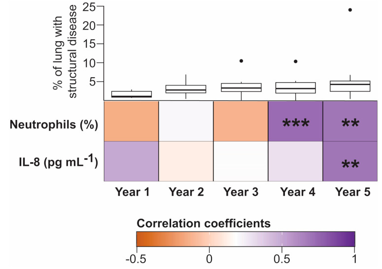
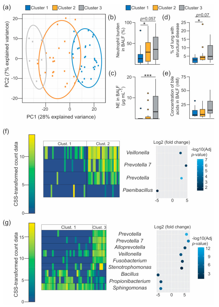
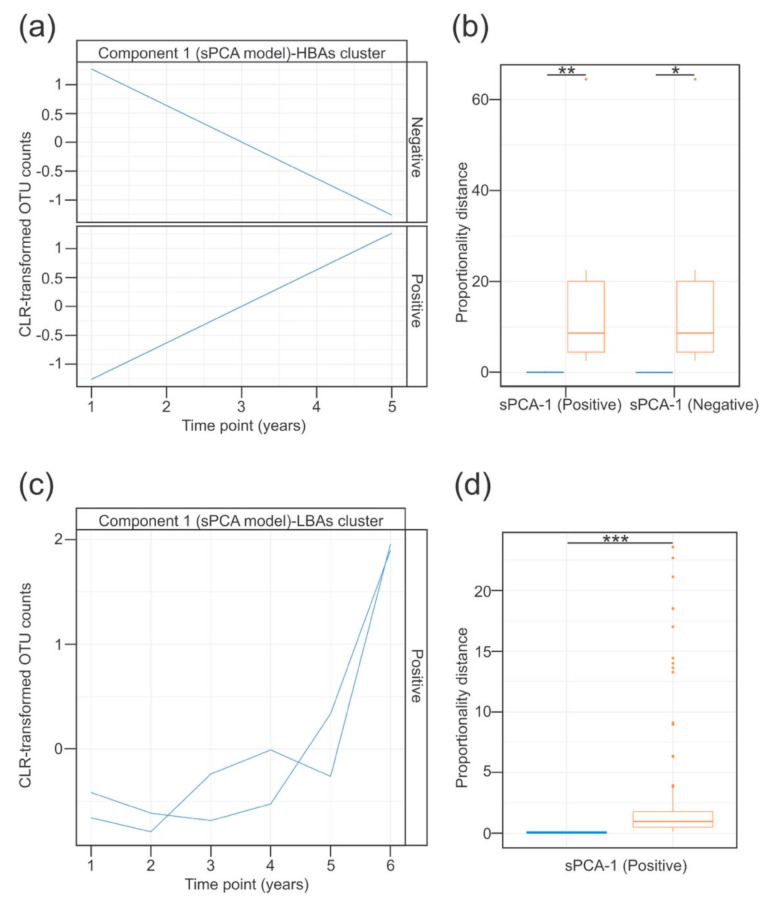
Cystic fibrosis (CF) is a congenital disorder resulting in a multisystemic impairment in ion homeostasis. The subsequent alteration of electrochemical gradients severely compromises the function of the airway epithelia. These functional changes are accompanied by recurrent cycles of inflammation-infection that progressively lead to pulmonary insufficiency. Recent developments have pointed to the existence of a gut-lung axis connection, which may modulate the progression of lung disease. Molecular signals governing the interplay between these two organs are therefore candidate molecules requiring further clinical evaluation as potential biomarkers. We demonstrate a temporal association between bile acid (BA) metabolites and inflammatory markers in bronchoalveolar lavage fluid (BALF) from clinically stable children with CF. By modelling the BALF-associated microbial communities, we demonstrate that profiles enriched in operational taxonomic units assigned to supraglottic taxa and opportunistic pathogens are closely associated with inflammatory biomarkers. Applying regression analyses, we also confirmed a linear link between BA concentration and pathogen abundance in BALF. Analysis of the time series data suggests that the continuous detection of BAs in BALF is linked to differential ecological succession trajectories of the lung microbiota. Our data provide further evidence supporting a role for BAs in the early pathogenesis and progression of CF lung disease.

Keywords: bile acids; cystic fibrosis; gut–lung axis; inflammation; lung microbiota.


Conflict of interest statement

The authors declare no conflict of interest. The funders had no role in the design of the study; in the collection, analyses, or interpretation of data; in the writing of the manuscript, or in the decision to publish the results.

Figures

Figure 1
Figure 1
Temporal associations between bile acids (BAs) and inflammatory markers in bronchoalveolar lavage fluid (BALF). The heatmap represents the Spearman rank-order correlation coefficients between total BA concentration (log10 transformed) and the indicated inflammatory biomarkers in BALF. Temporal co-occurrence is represented in samples obtained from one- to five-year-old cystic fibrosis (CF) subjects (Number of independent samples: year 1, n = 13; year 2, n = 14; year 3, n = 13; year 4, n = 16; year 5, n = 13). Boxplots in the top row represent the percentage of structural lung disease over the five-year period quantified using the PRAGMA protocol [36]. No significant differences in airway remodelling were observed in the context of Dunnett’s test between year 1 and each of the following four years. Asterisks indicate statistical significance for the indicated correlation per year after false discovery rate (FDR) correction. **, p < 0.01; ***, p < 0.001.
Figure 2
Figure 2
Inflammatory surrogates in BALF are linked to specific microbial assemblages. (a) Principal component analysis decomposition of the OTU counts transformed using the cumulative sum scaling (CSS) normalization method. Mean-centering was performed to better represent the direction of variability across group centroids. BALF-associated taxonomic profiles are projected onto the first two components of the model. Dots represent each BALF specimens, which are coloured accordingly with the Gaussian Mixture Model-based cluster membership (cluster 1, n = 29 BALF specimens; cluster 2, n = 22; cluster 3, n = 8). Ellipses represent the 86% confidence region. (be) Boxplots showing between-cluster comparisons of the indicated markers of disease progression (bd) and bile acid concentration (e) in BALF. Multiple comparisons were assessed for significance in the context of Dunnett’s test. *, p < 0.05; ***, p < 0.001. (f,g) Heatmaps (left) and bivariate dot plot (right) representing the CSS-normalised count data and the fold change (log2 scale) for the selected OTUs between the indicated clusters (Clust.), respectively. Differentially abundant features between clusters were estimated from a zero-inflated log-normal model as implemented in the fitFeatureModel method [20]. Features with a log2 (fold change) > |2| and an FDR-corrected (adjusted p-value) p-value < 0.05 are depicted. Colour legend for the heatmap (left), and the dot plot (right), represent the CSS-transformed count data and the –log10 (adjusted p-value) respectively. Each column in the heatmaps represents the profiles of individual BALF samples, and rows represent the indicated OTUs.
Figure 3
Figure 3
Longitudinal modelling of the BALF-associated microbial profiles in patients clustered based on BA detection patterns. (ad) Temporal profiles of the OTUs grouped using sPCA in the HBA (A) or LBA (C) patient clusters. Blue traces represent the modelled abundance (Centred Log Ratio (CLR)-transformed) of the OTUs selected in the sPCA model across time. Within each component, the selected OTU profiles were subclustered into positive or negative according to the direction of the correlation between each OTU profile, and the indicated latent variable of the sPCA model. (b,d) Association between the features assigned to the same cluster was evaluated using proportionality distances. Bar plot in (b) (for the HBA subset) shows the proportionality distance of features from the same cluster (blue), and the distance of OTUs inside that cluster with every feature in the other cluster (orange). Given that only one cluster was selected in the LBA group, the bar plot in (d) represents the proportionality distance between pairs of features from the cluster (blue), and between each feature in that cluster and the rest of features in the entire dataset (orange). In both cases, the intra-cluster distance was significantly lower than the distance outside the cluster, suggesting a strong association between the selected OTU profiles clustered together. *, p < 0.05; **, p < 0.01; ***, p < 0.001 in the context of the Wilcoxon test. Type I error rate was controlled with the false discovery rate method.

Similar articles

Cited by

References

    1. Riordan J.R., Rommens J.M., Kerem B., Alon N., Rozmahel R., Grzelczak Z., Zielenski J., Lok S., Plavsic N., Chou J.L., et al. Identification of the cystic fibrosis gene: Cloning and characterization of complementary DNA. Science. 1989;245:1066–1073. doi: 10.1126/science.2475911. - DOI - PubMed
    1. Elborn J.S. Cystic fibrosis. Lancet. 2016;388:2519–2531. doi: 10.1016/S0140-6736(16)00576-6. - DOI - PubMed
    1. Kreda S.M., Davis C.W., Rose M.C. CFTR, mucins, and mucus obstruction in cystic fibrosis. Cold Spring Harb. Perspect. Med. 2012;2:a009589. doi: 10.1101/cshperspect.a009589. - DOI - PMC - PubMed
    1. Ranganathan S.C., Hall G.L., Sly P.D., Stick S.M., Douglas T.A., Australian Respiratory Early Surveillance Team for Cystic, F Early Lung Disease in Infants and Preschool Children with Cystic Fibrosis. What Have We Learned and What Should We Do about It? Am. J. Respir. Crit. Care Med. 2017;195:1567–1575. doi: 10.1164/rccm.201606-1107CI. - DOI - PMC - PubMed
    1. Sly P.D., Gangell C.L., Chen L., Ware R.S., Ranganathan S., Mott L.S., Murray C.P., Stick S.M., Investigators A.C. Risk factors for bronchiectasis in children with cystic fibrosis. N. Engl. J. Med. 2013;368:1963–1970. doi: 10.1056/NEJMoa1301725. - DOI - PubMed

LinkOut - more resources