. 2020 Nov 6;25(21):5169.
doi: 10.3390/molecules25215169.

AGS3 and Gαi3 Are Concomitantly Upregulated as Part of the Spindle Orientation Complex during Differentiation of Human Neural Progenitor Cells

Affiliations

AGS3 and Gαi3 Are Concomitantly Upregulated as Part of the Spindle Orientation Complex during Differentiation of Human Neural Progenitor Cells

Jackson L K Yip et al. Molecules. .

Abstract

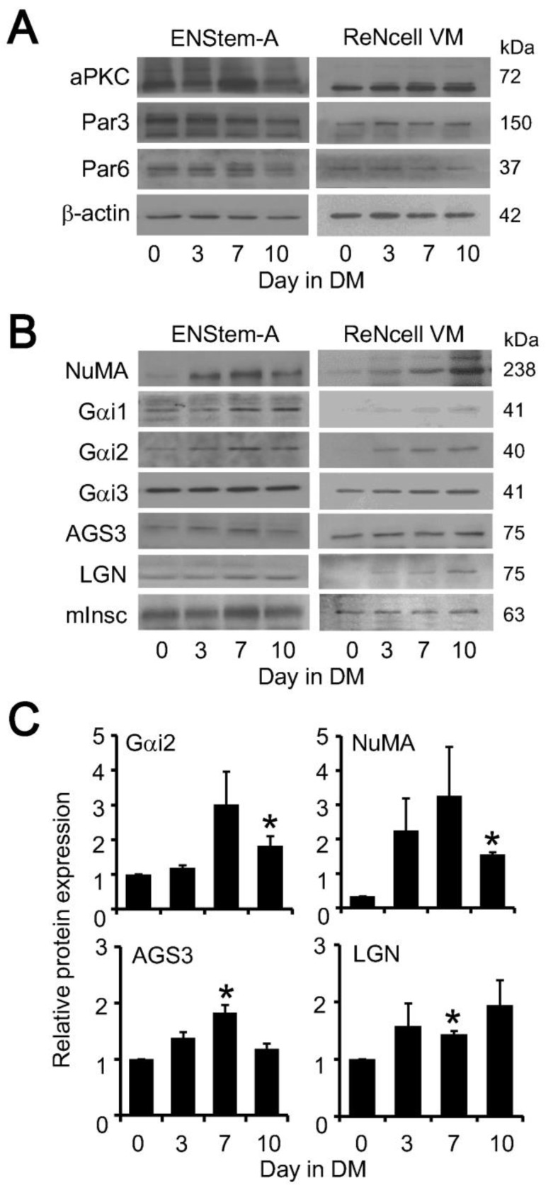
Adult neurogenesis is modulated by many Gi-coupled receptors but the precise mechanism remains elusive. A key step for maintaining the population of neural stem cells in the adult is asymmetric cell division (ACD), a process which entails the formation of two evolutionarily conserved protein complexes that establish the cell polarity and spindle orientation. Since ACD is extremely difficult to monitor in stratified tissues such as the vertebrate brain, we employed human neural progenitor cell lines to examine the regulation of the polarity and spindle orientation complexes during neuronal differentiation. Several components of the spindle orientation complex, but not those of the polarity complex, were upregulated upon differentiation of ENStem-A and ReNcell VM neural progenitor cells. Increased expression of nuclear mitotic apparatus (NuMA), Gαi subunit, and activators of G protein signaling (AGS3 and LGN) coincided with the appearance of a neuronal marker (β-III tubulin) and the concomitant loss of neural progenitor cell markers (nestin and Sox-2). Co-immunoprecipitation assays demonstrated that both Gαi3 and NuMA were associated with AGS3 in differentiated ENStem-A cells. Interestingly, AGS3 appeared to preferentially interact with Gαi3 in ENStem-A cells, and this specificity for Gαi3 was recapitulated in co-immunoprecipitation experiments using HEK293 cells transiently overexpressing GST-tagged AGS3 and different Gαi subunits. Moreover, the binding of Gαi3 to AGS3 was suppressed by GTPγS and pertussis toxin. Disruption of AGS3/Gαi3 interaction by pertussis toxin indicates that AGS3 may recognize the same site on the Gα subunit as G protein-coupled receptors. Regulatory mechanisms controlling the formation of spindle orientation complex may provide novel means to manipulate ACD which in turn may have an impact on neurogenesis.

Keywords: AGS3; G protein; LGN; NuMA; Ric-8A; neurogenesis.


Conflict of interest statement

The authors declare no conflict of interest.

Figures

Figure 1
Figure 1
Differentiation of human neural progenitor cells. (A) ENStem-A cells were induced to undergo differentiation at passage number 6 while ReNcell VM cells were induced at passage number 9. Cell lysates were collected at Day 0, Day 3, Day 7 and Day 10 of differentiation and subjected to Western blot analysis using anti-Nestin, anti-Sox-2, anti-β-III tubulin and anti-β-actin antisera. Data shown represent one of three or more sets of immunoblots; other sets yielded similar results. (B) ENStem-A cells were fixed by 4% paraformaldehyde at Day 0 and Day 7 of differentiation. Cell nuclei were stained with DAPI (4’,6-diamidino-2-phenylindole) while Sox-2 and, β-III tubulin were identified by specific antisera and fluorescent secondary antibodies. Images were obtained with a Zeiss LSM700 confocal microscope. (C) ReNcell VM cells were processed as in B except anti-nestin was used instead of anti-Sox-2 for assessing pluripotency. Scale bar indicates 10 μm.
Figure 2
Figure 2
Upregulation of spindle orientation proteins in human neural progenitor cells during differentiation. ENStem-A and ReNcell VM cells were induced to undergo differentiation. Cell lysates were collected at Day 0, Day 3, Day 7 and Day 10 of differentiation. (A) Expression of polarity proteins were determined by Western blot analysis using anti-atypical protein kinase C (aPKC), anti-partition defective 3 (Par3), anti-Par6 and anti-β-actin antisera. (B) Expression of spindle orientation proteins were assessed by Western blot analysis using anti-nuclear mitotic apparatus (NuMA), anti-Gαi1, anti-Gαi2, anti-Gαi3, anti-AGS3, anti-LGN and anti-inscuteable antisera; please see β-actin immunoreactivity in panel A for reference protein expression. Data shown represent one of three or more sets of immunoblots; other sets yielded similar results. (C) Quantitative analysis of the expression of Gαi2, NuMA, AGS3 and LGN using the ImageJ software. Data were expressed as the mean ± S.E. of at least three independent sets of experiments. The probability of an observed difference being a coincidence was evaluated by Dunnett t test. Differences at values of p < 0.05 were considered significant (* p < 0.05).
Figure 3
Figure 3
Co-localization and complex formation by upregulated spindle orientation proteins during differentiation of ENStem-A cells. (A) ENStem-A cells were fixed by 4% paraformaldehyde at Day 0 and Day 7 of differentiation. Cell nuclei were stained with DAPI and the presence of NuMA, LGN, and AGS3 detected by fluorescence staining using specific antisera. Images were obtained with a Zeiss LSM700 confocal microscope. Scale bar indicates 10 μm. (B) Images of LGN and NuMA at Day 7 were merged (bottom panel) to illustrate their co-localization. Scale bar indicates 10 μm. (C) ENStem-A cells were induced to undergo differentiation and total RNA was extracted using RNeasy Mini kit (Qiagen) at Day 0, Day 3, Day 7 and Day 10. Each RNA sample was then subjected to reverse transcription to generate cDNA followed by PCR amplification with primers corresponding to Gαi1, Gαi2, Gαi3, AGS3, LGN, NuMA and GAPDH. (D) ENStem-A cells were induced to undergo differentiation and cell lysates were collected at Day 0 and Day 7 of differentiation. Samples were incubated with anti-AGS3 antisera and then immunoprecipitated using protein G agarose. Presence of Gαi2, Gαi3 and NuMA in the immunoprecipitates (IP) and cell lysates were detected by Western blotting with specific antisera.
Figure 4
Figure 4
Interaction of Gαi with components of the spindle orientation complex in HEK293 cells. (A) HEK293 cells were transiently transfected with vector (pcDNA3.1), or co-transfected with GST-tagged AGS3 and Gαi1, Gαi2 or Gαi3. Samples were incubated with anti-GST and then immunoprecipitated using protein G agarose. Presence of Gαi1, Gαi2, Gαi3 and GST-tagged AGS3 were analyzed by Western blotting with specific antisera. (B) HEK293 cells were transiently transfected with vector (pcDNA3.1), or co-transfected with GST-tagged AGS3 or GFP tagged LGN and Gαi2 or Gαi3. One day after transfection, cells was treated with or without pertussis toxin (PTX) for 16 h. Cell lysates were subjected to treatment with GDPβS and GTPγS (100 μM; 6 h at 4 °C). Samples were subjected to immunoprecipitation with either anti-GST or anti-GFP antisera and protein G agarose. Gαi subunits in the immunoprecipitates were detected by Western blot analysis. (C) HEK293 cells transiently co-expressing HA-tagged NuMA and Gαi3 were subjected to co-immunoprecipitation with anti-HA agarose and Western blotting as in B. (D) HEK293 cells were treated as in C except that HA-tagged Ric-8A was transfected and probed instead of HA-tagged NuMA.
Figure 5
Figure 5
Crystal structures of Gαi-GDP in complex with GoLoco motif or Gβγ subunits. (A) Ribbon diagram of the Gαi-GDP in complex with GoLoco motif crystal structure in two views related by a 90° rotation about the vertical axis. The model of a full length Gα structure (residues 5-354) was constructed by combining published crystal structures of Gαi subunits (PDB entries 4G5R and 1GP2). Selected amino acids suggested to participate in GoLoco motif interaction (Tyr69, Val72, Ser75, Gln79, Val179 and Thr181; Jia et al., 2012) are highlighted in orange. Gαi and GoLoco peptide are shown in light gray and red, respectively. The PTX ADP-ribosylation site (Cys351) is highlighted in magenta. GDP is shown in the ball-and-stick model (green). (B) Ribbon diagram of the Gαi-GDP in complex with Gβγ subunits crystal structure. Gβ and Gγ subunits are shown in blue and lime. The color scheme of Gαi is the same as in A.

Similar articles

Cited by

References

    1. Taverna E., Gotz M., Huttner W.B. The cell biology of neurogenesis: Toward an understanding of the development and evolution of the neocortex. Annu. Rev. Cell Dev. Biol. 2014;30:465–502. doi: 10.1146/annurev-cellbio-101011-155801. - DOI - PubMed
    1. Shen Q., Zhong W., Jan Y.N., Temple S. Asymmetric Numb distribution is critical for asymmetric cell division of mouse cerebral cortical stem cells and neuroblasts. Development. 2002;129:4843–4853. - PubMed
    1. Shitamukai A., Matsuzaki F. Control of asymmetric cell division of mammalian neural progenitors. Dev. Growth Differ. 2012;54:277–286. doi: 10.1111/j.1440-169X.2012.01345.x. - DOI - PubMed
    1. Suzuki A., Ohno S. The PAR-aPKC system: Lessons in polarity. J. Cell Sci. 2006;119:979–987. doi: 10.1242/jcs.02898. - DOI - PubMed
    1. Bowman S.K., Neumuller R.A., Novatchkova M., Du Q., Knoblich J.A. The Drosophila NuMA homolog Mud regulates spindle orientation in asymmetric cell division. Dev. Cell. 2006;10:731–742. doi: 10.1016/j.devcel.2006.05.005. - DOI - PubMed

MeSH terms

Substances