. 2021 Oct;17(10):2842-2855.
doi: 10.1080/15548627.2020.1847444. Epub 2020 Nov 23.

HPV sensitizes OPSCC cells to cisplatin-induced apoptosis by inhibiting autophagy through E7-mediated degradation of AMBRA1

Affiliations

HPV sensitizes OPSCC cells to cisplatin-induced apoptosis by inhibiting autophagy through E7-mediated degradation of AMBRA1

Manuela Antonioli et al. Autophagy. 2021 Oct.

Abstract

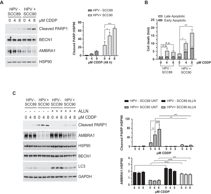
Oropharyngeal squamous cell carcinoma (OPSCC) is an increasing world health problem with a more favorable prognosis for patients with human papillomavirus (HPV)-positive tumors compared to those with HPV-negative OPSCC. How HPV confers a less aggressive phenotype, however, remains undefined. We demonstrated that HPV-positive OPSCC cells display reduced macroautophagy/autophagy activity, mediated by the ability of HPV-E7 to interact with AMBRA1, to compete with its binding to BECN1 and to trigger its calpain-dependent degradation. Moreover, we have shown that AMBRA1 downregulation and pharmacological inhibition of autophagy sensitized HPV-negative OPSCC cells to the cytotoxic effects of cisplatin. Importantly, semi-quantitative immunohistochemical analysis in primary OPSCCs confirmed that AMBRA1 expression is reduced in HPV-positive compared to HPV-negative tumors. Collectively, these data identify AMBRA1 as a key target of HPV to impair autophagy and propose the targeting of autophagy as a viable therapeutic strategy to improve treatment response of HPV-negative OPSCC.Abbreviations: AMBRA1: autophagy and beclin 1 regulator 1; CDDP: cisplatin (CDDP); FFPE: formalin-fixed paraffin-embedded (FFPE); HNC: head and neck cancers (HNC); HPV: human papillomavirus (HPV); hrHPV: high risk human papillomavirus (hrHPV); OCSCC: oral cavity squamous carcinomas (OCSSC); OPSCC: oropharyngeal squamous cell carcinoma (OPSCC); OS: overall survival (OS); qPCR: quantitative polymerase chain reaction; RB1: RB transcriptional corepressor 1; ROC: receiver operating characteristic curve (ROC).

Keywords: AMBRA1; autophagy; calpains; hpv-E7; oropharyngeal squamous cell carcinoma.


Conflict of interest statement

Dr Rob Ellis, Dr Marie Labus and Prof. Penny Lovat are directors of AMLo Biosciences Ltd. The other authors declare that they have no conflict of interest.

Figures

Figure 1.
Figure 1.
Autophagy is impaired in HPV-positive OPSCC. (A) LC3 and SQSTM1/p62 western blotting analysis of HPV-negative (SCC89 and SCC25) and HPV-positive (SCC90 and SCC154) starved cells. (B) Analysis of the autophagic flux (LC3 and SQSTM1/p62 blot) of HPV-negative and -positive cells deprived of nutrients without bafilomycin A1 for 1 h at 5 nM (n = 4, mean ± SEM. ANOVA 2-way test, *P < 0.05, **P < 0.005, ****P < 0.0005). (C) Analysis of LC3 and LAMP1 colocalization expressed as the number of dots per cell. HPV-negative SCC89 and HPV-positive SCC90 cells were subjected to nutrient deprivation for 2 h (starvation) and treated or not with the lysosomal inhibitor bafilomycin A1 (bafA1). Numbers under the x-axis represent the fold of change compared to control of each cell line (n = 30 cells, mean ± SEM. ANOVA 2-way test, *P < 0.05, **P < 0.005). Statistical analysis are limited to untreated/bafA1-treated paired samples as an indication of autophagy flux modulation
Figure 2.
Figure 2.
Relationship between HPV status and AMBRA1 expression in OPSCC. (A) Western blotting analysis of several autophagic proteins comparing HPV-negative SCC89 and HPV-positive SCC90 starved cells. Lower graph: densitometric analysis of AMBRA1 protein levels (n = 3, mean ± SEM. ANOVA 2-way test, ****P < 0.00005). (B) Overall survival rates of Newcastle OPSCC cohort (83) determined by Kaplan-Meier analysis and compared by Log-Rank test in HPV positive versus HPV negative OPSCC samples (OS = 63% versus 32.7%; P = 0.0012 HR 2.87 (95% CI 1.54–5.35). (C) Representative photomicrographs of AMBRA1 expression in a cohort of 83 Newcastle OPSCC TMA i. AMBRA1 H-Score = 300 ii. H-score = 180 iii. H-Score = 120 iv. H-score = 20. Scale bars = 300 µm. (D) Median tumoral expression of AMBRA1 in the Newcastle cohort of 45 HPV negative or 38 HPV positive OPSCCs revealing significantly greater levels of expression (h score) in HPV negative OPSCC (H-score = 150 (IQR 100)) compared to HPV positive OPSCC (H-score = 100 (IQR 117.75). Mann-Whitney P = 0.049)
Figure 3.
Figure 3.
HPV16 E7 promotes a calpain-mediated degradation of AMBRA1. (A) Quantitative PCR of AMBRA1 mRNA comparing HPV-negative SCC89 and HPV-positive SCC90 cells. (B) Western blot analysis of AMBRA1 protein levels in HPV-negative SCC89 and HPV-positive SCC90 cells, where proteasomal, calpains or lysosomal activities were blocked using MG132 at 5 mM, ALLN at 10 mM and bafilomycin A1 at 5 nM for 5 h, respectively. Lower panel shows the densitometric analysis of 3 independent experiment reported as the fold of changes compared the each untreated cell line (mean ± SEM, ANOVA 2-way test, *P < 0.05, **P < 0.005, ***P < 0.005). (C) AMBRA1 protein levels following the expression of HA/FLAG-tagged HPV16 E7 in 293 T cells (n = 3), (D) in presence or absence of ALLN at 10 mM for 5 h (n = 3)
Figure 4.
Figure 4.
HPV16 E7 interacts with AMBRA1 to compete with BECN1 interaction and to promote CAPN2 association. (A) Immunoprecipitation of expressed HA/FLAG-tagged HPV16 E7 in 293 T cells revealed for AMBRA1, Rb and BECN1 interaction by western blot. (B) Immunoprecipitation of endogenous HPV16 E7 from HPV-positive OPSCC protein extracts probed with anti-AMBRA1 antibody. (C) Schematic representation of different AMBRA1 deletion mutants. (D) Immunoprecipitation of different MYC-tagged AMBRA1 deletion mutants in presence of expressing HA/FLAG-tagged HPV16 E7 in 293 T cells. (E) Immunoprecipitation of BECN1 using a specific antibody in 293 T cells expressing both Myc-tagged AMBRA1 and HA/FLAG-tagged E7. (F) Analysis of AMBRA1-BECN1 interaction in presence or not of HPV16 E7 performed by immunoprecipitation of endogenous AMBRA1 revealed for BECN1 by western blotting, the lower panel represents the amount of BECN1 co-eluted and normalized on immunoprecipitated AMBRA1 (n = 3, mean ± SEM. Two-tailed Student’s t-test, ****P < 0.00005). (G) Immunoprecipitation of FLAG-tagged AMBRA1 expressed in 293 T cells revealed with anti-CAPN2 antibody. (H) Immunoprecipitation FLAG-tagged AMBRA1 expressing 293 T in presence or not of HA-tagged HPV16 E7, CAPN2 interaction was detected by western blot. The right panel represents the amount of CAPN2 co-eluted and normalized on immunoprecipitated AMBRA1 (n = 4, mean ± SEM. Two-tailed Student’s t-test, *P < 0.05)
Figure 5.
Figure 5.
Calpains are responsible for the increased susceptibility of HPV-positive OPSCC cells to Cisplatin-induced cell death. (A) Analysis of cell death comparing HPV-negative and positive OPSCC cells in response Cisplatin (CDDP) treatment for 4 or 8 μM (48 h) analyzed by western blotting of cleaved PARP1, (B) and evaluating PI/Annexin V incorporation by FACS (n = 3, mean ± SEM. ANOVA 2-way test, *P < 0.05 **P < 0.005). (C) Cleaved PARP1 western blotting of HPV-negative and positive OPSCC cells treated with increasing amount of Cisplatin in presence or absence of ALLN and relative densitometric analysis; several autophagic proteins were evaluated as control (n = 4, mean ± SEM. ANOVA 2-way test, P** P < 0.005 *** P < 0.0005)
Figure 6.
Figure 6.
Autophagy is responsible for cisplatin-resistance of HPV-negative OPSCC cells. (A) Lentiviral infected HPV-negative OPSCC cells for shRNA of AMBRA1 using two different vectors and (B) stable AMBRA1 KO HPV-negative OPSCC cells, tested for Cisplatin-induced apoptosis by western blotting, and (C) by FACS. (D) HPV-negative OPSCC cells infected with lentivirus for BECN1 shRNA, tested for PARP1 cleavage by western blotting. (E-G) HPV-negative OPSCC cells where autophagy was inhibited or not using (E) 3-MA at 2.5 mM or, (F) and (G) PIK-III at 2.5 μM, and cell death stimulated by cisplatin treatment for 48 h. n = 3, mean ± SEM. ANOVA 2-way test, * P < 0.05, ** P < 0.005, **** P < 0.00005

References

    1. Amin MB, Greene FL, Edge SB, et al. The eighth edition AJCC cancer staging manual: continuing to build a bridge from a population-based to a more “personalized” approach to cancer staging. CA Cancer J Clin [Internet] 2017. [cited 2019 Apr 4]; 67(2):93–99. Available from: http://www.ncbi.nlm.nih.gov/pubmed/28094848 - PubMed
    1. IARC working group on the evaluation of carcinogenic risks to humans. Biological agents. Volume 100 B. A review of human carcinogens. IARC Monogr Eval Carcinog Risks Hum [Internet] 2012. [cited 2019 Aug 5]; 100:1–441. Available from: http://www.ncbi.nlm.nih.gov/pubmed/23189750 - PMC - PubMed
    1. Vescovo T, Pagni B, Piacentini M, et al. Regulation of autophagy in cells infected with oncogenic human viruses and its impact on cancer development. Front Cell Dev Biol. 2020;8:47. - PMC - PubMed
    1. Schiffman M, Doorbar J, Wentzensen N, et al. Carcinogenic human papillomavirus infection. Nat Rev Dis Prim. [Internet] 2016. [cited 2019 Jul 23]; 2:16086. Available from: http://www.nature.com/articles/nrdp201686 - PubMed
    1. Darnell GA, Schroder WA, Antalis TM, et al. Human Papillomavirus E7 requires the protease calpain to degrade the retinoblastoma protein. J Biol Chem [Internet] 2007. [cited 2019 Apr 4]; 282(52):37492–37500. Available from: http://www.ncbi.nlm.nih.gov/pubmed/17977825 - PubMed

Publication types

MeSH terms