. 2020 Nov 10;12(1):29.
doi: 10.1186/s11689-020-09332-3.

A small-molecule screen reveals novel modulators of MeCP2 and X-chromosome inactivation maintenance

Affiliations

A small-molecule screen reveals novel modulators of MeCP2 and X-chromosome inactivation maintenance

Hyeong-Min Lee et al. J Neurodev Disord. .

Abstract

Background: Rett syndrome (RTT) is a neurodevelopmental disorder caused by mutations in the X-linked methyl-CpG binding protein 2 (MeCP2) gene. While MeCP2 mutations are lethal in most males, females survive birth but show severe neurological defects. Because X-chromosome inactivation (XCI) is a random process, approximately 50% of the cells silence the wild-type (WT) copy of the MeCP2 gene. Thus, reactivating the silent WT copy of MeCP2 could provide therapeutic intervention for RTT.

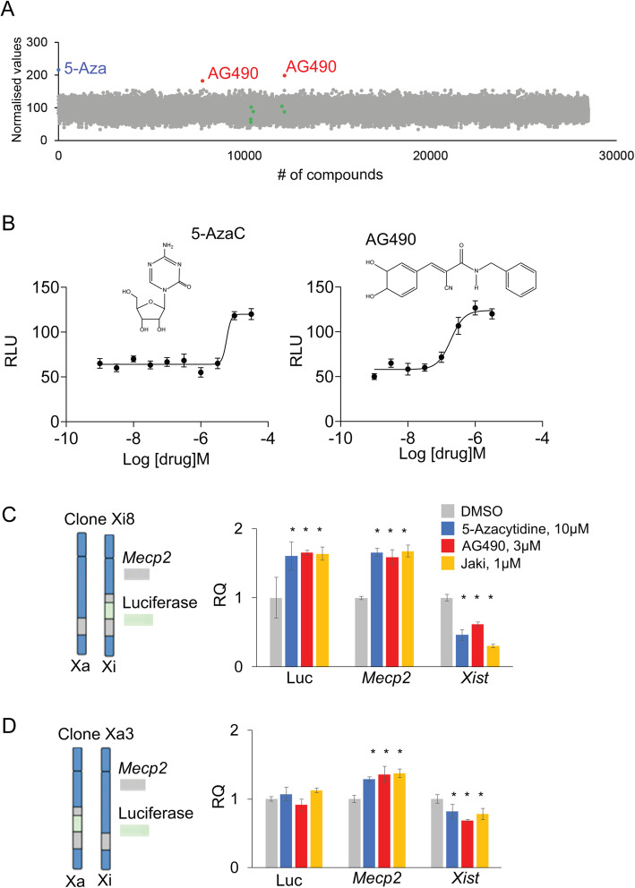
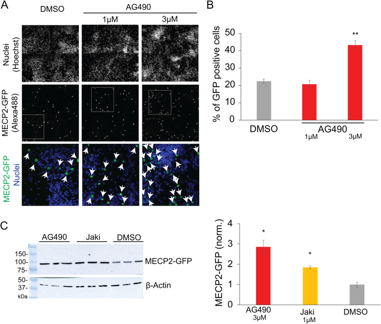
Methods: Toward this goal, we screened ~ 28,000 small-molecule compounds from several libraries using a MeCP2-luciferase reporter cell line and cortical neurons from a MeCP2-EGFP mouse model. We used gain/increase of luminescence or fluorescence as a readout of MeCP2 reactivation and tested the efficacy of these drugs under different drug regimens, conditions, and cellular contexts.

Results: We identified inhibitors of the JAK/STAT pathway as XCI-reactivating agents, both by in vitro and ex vivo assays. In particular, we show that AG-490, a Janus Kinase 2 (JAK2) kinase inhibitor, and Jaki, a pan JAK/STAT inhibitor, are capable of reactivating MeCP2 from the inactive X chromosome, in different cellular contexts.

Conclusions: Our results suggest that inhibition of the JAK/STAT pathway is a new potential pathway to reinstate MeCP2 gene expression as an efficient RTT treatment.

Keywords: AG490; JAK/STAT; Janus Kinase; Janus Kinase inhibitors; MeCP2; PI3K/ATK pathways; Rett syndrome; X-chromosome inactivation.


Conflict of interest statement

The authors declare no competing interests.

Figures

Fig. 1
Fig. 1
Luminescence-based screen identifies AG490 and Jaki as potential reactivators of genes on the inactive X chromosome. a Dot plot indicating the percentage of luciferase activation per drug using the Xi8 fibroblast cell line reporter for MeCP2 activation over the control (normalized values; see the “Material and methods” section). In red, we show AG490, in blue 5-Aza, and in green compounds previously reported to reactivate MeCP2 using different protocols (LY294002, OSU-03012, MLN8237, BX-912, K02288, VX-680; see the “Introduction” and the “Results” sections for more details). b Top: Chemical structure of 5-azacytidine (5-Aza), a known reactivator of MeCP2, and AG490. Bottom: Dose response of 3-day treatment of 5-Aza or AG490 in Xi8 fibroblast cells (n = 3, one representative graph shown here from three independent assays). c Left: Xi8 clone showing MeCP2 and MeCP2-luciferase genes on the inactive chromosome. Right: qRT-PCR data showing the effect of the indicated drug treatments on luciferase (Luc), Mecp2, and Xist mRNA levels; mean (± SEM) are shown, *statistical significance, p ≤ 0.05 (n = 3 per compounds, the average of three biological replicates are shown). d Left: Xa3 clone showing MeCP2 and MeCP2-luciferase genes on the active chromosome, respectively. Right: qRT-PCR data showing the effect of the indicated drug treatments on luciferase (Luc), Mecp2, and Xist mRNA levels; mean (± SEM) are shown, *statistical significance, p ≤ 0.05 (n = 3 per compounds, the average of three biological replicates are shown)
Fig. 2
Fig. 2
AG490 effectively upregulates MeCP2 in the cultured cortical neurons. a Representative fluorescent immunostaining images of cortical neurons after treating with AG490 or DMSO vehicle twice for 3 days each. The top row shows labeling of nuclei, and the middle row shows GFP labeling (of MeCP2-GFP). The bottom row shows the zoom-in of nuclear and MeCP2-GFP labeling from the boxed area depicted in the middle row. Arrows indicate GFP-positive cells. b Quantitative analysis of the number of GFP-positive cells (**p ≤ 0.01) (n = 4 per dose, the average of three biological replicates are shown). c GFP western blot analysis (left) and WB quantification (right) show MeCP2 upregulation in AG490- and Jaki-treated cells, *statistical significance, p ≤ 0.05 (n = 3 per compounds, the average of two biological replicates are shown)
Fig. 3
Fig. 3
AG490 upregulates MeCP2 in different cellular contexts. a qRT-PCR analysis of AG490 treatments in a humanized Xi-containing line (THX88) is shown. Dots indicate individual experimental points, samples are color-coded (see legend), and concentrations are indicated. ***Statistical significance, p ≤ 0.001, one-way ANOVA. The experiments were done using 2 (5-Aza), 3 (Jaki), and 4 (AG490) biological replicates, using 2 to 3 technical replicates per experiment. Horizontal lines displayed the median of the samples. Gapdh was used as an internal normalization control. b Survival analysis of H4SV cells in HAT-selecting medium in the presence or absence of Xi-reactivating compounds. Fold changes (FC, A.U) of the total number of colonies for control (DMSO) and Xi-reactivating drugs are shown. Bars indicate the standard deviation (SD). *Statistical significance, p ≤ 0.05. The average of three biological replicates are shown. Representative plates are shown below. c qRT-PCR data showing the effect of the indicated drug treatments on Mecp2, Hprt, and Xist mRNA levels; mean (± SD) are shown, *statistical significance, p ≤ 0.05 (n = 3 per compounds, the average of three biological replicates are shown)

References

    1. Amir RE, Van den Veyver IB, Wan M, Tran CQ, Francke U, Zoghbi HY. Rett syndrome is caused by mutations in X-linked MECP2, encoding methyl-CpG-binding protein 2. Nature Genetics. 1999;23(2):185–188. doi: 10.1038/13810. - DOI - PubMed
    1. Degano AL, Pasterkamp RJ, Ronnett GV. MeCP2 deficiency disrupts axonal guidance, fasciculation, and targeting by altering Semaphorin 3F function. Mol Cell Neurosci. 2009;42(3):243–254. doi: 10.1016/j.mcn.2009.07.009. - DOI - PMC - PubMed
    1. Matarazzo V, Cohen D, Palmer AM, Simpson PJ, Khokhar B, Pan SJ, Ronnett GV. The transcriptional repressor Mecp2 regulates terminal neuronal differentiation. Mol Cell Neurosci. 2004;27(1):44–58. doi: 10.1016/j.mcn.2004.05.005. - DOI - PubMed
    1. Noutel J, Hong YK, Leu B, Kang E, Chen C. Experience-dependent retinogeniculate synapse remodeling is abnormal in MeCP2-deficient mice. Neuron. 2011;70(1):35–42. doi: 10.1016/j.neuron.2011.03.001. - DOI - PMC - PubMed
    1. Cohen S, Gabel HW, Hemberg M, Hutchinson AN, Sadacca LA, Ebert DH, Harmin DA, Greenberg RS, Verdine VK, Zhou Z, et al. Genome-wide activity-dependent MeCP2 phosphorylation regulates nervous system development and function. Neuron. 2011;72(1):72–85. doi: 10.1016/j.neuron.2011.08.022. - DOI - PMC - PubMed

Publication types

Substances