. 2020 Nov 10;11(6):e02470-20.
doi: 10.1128/mBio.02470-20.

Magnesium Sensing Regulates Intestinal Colonization of Enterohemorrhagic Escherichia coli O157:H7

Affiliations

Magnesium Sensing Regulates Intestinal Colonization of Enterohemorrhagic Escherichia coli O157:H7

Yutao Liu et al. mBio. .

Abstract

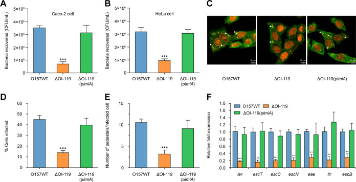
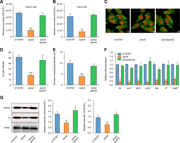
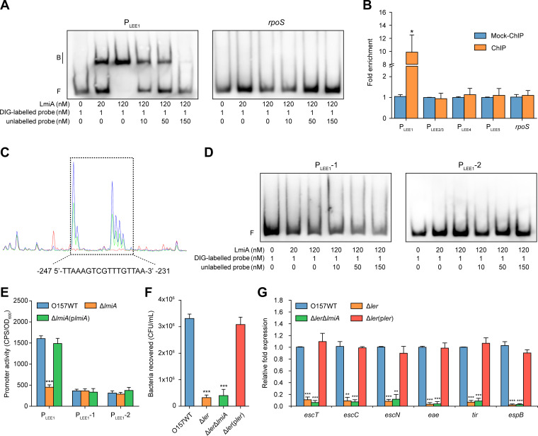
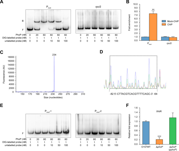
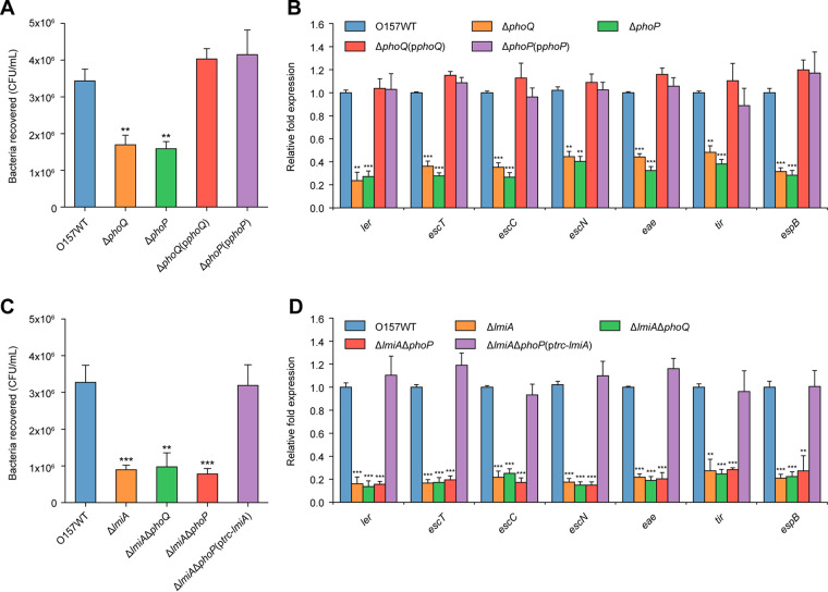
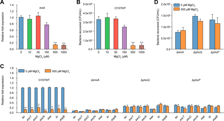
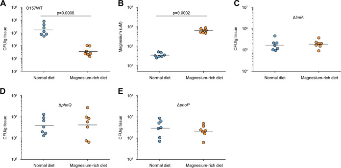
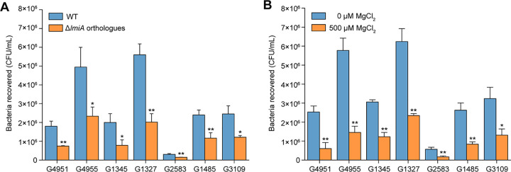
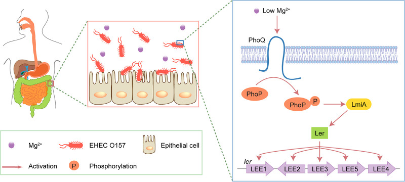
The large intestinal pathogen enterohemorrhagic Escherichia coli (EHEC) O157:H7 detects host cues to regulate virulence gene expression during colonization and infection. However, virulence regulatory mechanisms of EHEC O157:H7 in the human large intestine are not fully understood. Herein, we identified a virulence-regulating pathway where the PhoQ/PhoP two-component regulatory system senses low magnesium levels and signals to the O island 119-encoded Z4267 (LmiA; low magnesium-induced regulator A), directly activating loci of enterocyte effacement genes to promote EHEC O157:H7 adherence in the large intestine. Disruption of this pathway significantly decreased EHEC O157:H7 adherence in the mouse intestinal tract. Moreover, feeding mice a magnesium-rich diet significantly reduced EHEC O157:H7 adherence in vivo This LmiA-mediated virulence regulatory pathway is also conserved among several EHEC and enteropathogenic E. coli serotypes; therefore, our findings support the use of magnesium as a dietary supplement and provide greater insights into the dietary cues that can prevent enteric infections.IMPORTANCE Sensing specific gut metabolites is an important strategy for inducing crucial virulence programs by enterohemorrhagic Escherichia coli (EHEC) O157:H7 during colonization and infection. Here, we identified a virulence-regulating pathway wherein the PhoQ/PhoP two-component regulatory system signals to the O island 119-encoded low magnesium-induced regulator A (LmiA), which, in turn, activates locus of enterocyte effacement (LEE) genes to promote EHEC O157:H7 adherence in the low-magnesium conditions of the large intestine. This regulatory pathway is widely present in a range of EHEC and enteropathogenic E. coli (EPEC) serotypes. Disruption of this pathway significantly decreased EHEC O157:H7 adherence in the mouse intestinal tract. Moreover, mice fed a magnesium-rich diet showed significantly reduced EHEC O157:H7 adherence in vivo, indicating that magnesium may help in preventing EHEC and EPEC infection in humans.

Keywords: Z4267; bacterial adherence; gene regulation; locus of enterocyte effacement (LEE); magnesium; virulence.


Figures

FIG 1
FIG 1
OI-119 is required for EHEC O157 adherence capacity and LEE gene expression. (A) Adherence capacity of O157 WT, ΔOI-119, and ΔOI-119(plmiA) to Caco-2 cells. (B) Adherence capacity of O157 WT, ΔOI-119, and ΔOI-119(plmiA) to HeLa cells. (C) FAS assay of HeLa cells infected with O157 WT, ΔOI-119, and ΔOI-119(plmiA). HeLa nuclei and bacteria were stained with propidium iodide (red), and HeLa cell actin cytoskeleton was stained with FITC-phalloidin (green). Pedestals are observed as green punctate structures associated with bacterial cells and are indicated by arrowheads. Scale bar, 10 μm. (D and E) FAS assay quantification of the percentage of HeLa cells infected (D) and the number of pedestals per infected cell (E). n = 300 cells per strain. (F) qRT-PCR of LEE gene expression in O157 WT, ΔOI-119, and ΔOI-119(plmiA). O157 WT, EHEC O157 wild-type strain; ΔOI-119, OI-119 mutant; ΔOI-119(plmiA), OI-119 mutant complemented with lmiA. Data are presented as the means ± SD; n = 3. **, P ≤ 0.01; ***, P ≤ 0.001 (Student's t test).
FIG 2
FIG 2
LmiA is a positive virulence regulator of EHEC O157. (A) Adhesion of O157 WT, ΔlmiA, and ΔlmiA(plmiA) to Caco-2 cells. (B) Adhesion of O157 WT, ΔlmiA, and ΔlmiA(plmiA) to HeLa cells. (C) FAS assay of HeLa cells infected with O157 WT, ΔlmiA, and ΔlmiA(plmiA). HeLa nuclei and bacteria were stained with propidium iodide (red), and HeLa cell actin cytoskeleton was stained with FITC-phalloidin (green). Pedestals are observed as green punctate structures associated with bacterial cells and are indicated by arrowheads. Scale bar, 10 μm. (D and E) FAS assay quantification of the percentage of HeLa cells infected (D) and the number of pedestals per infected cell (E). n = 300 cells per strain. (F) qRT-PCR of LEE gene expression changes in O157 WT, ΔlmiA, and ΔlmiA(plmiA). (G) Representative Western blot images and quantitative analysis of intimin and its receptor Tir in O157 WT, ΔlmiA, and ΔlmiA(plmiA). Protein levels were semiquantified using ImageJ. The relative intensity is shown as the ratio of the signal intensity of intimin or Tir to GroEL (loading control). O157 WT, EHEC O157 wild-type strain; ΔlmiA, lmiA mutant; ΔlmiA(plmiA), lmiA mutant complemented with lmiA. Data are presented as the means ± SD; n = 3. *, P ≤ 0.05; **, P ≤ 0.01; ***, P ≤ 0.001 (Student's t test).
FIG 3
FIG 3
LmiA mediates the expression of EHEC O157 LEE genes via Ler. (A) Gel mobility shift and competition assays of LmiA with the promoter region of LEE1 and rpoS (negative control). Positions of the bound (denoted with “B”) and free (denoted with “F”) probes are shown on the left, and the concentrations of the probe and purified LmiA are indicated at the bottom of each lane. (B) Fold enrichment of the promoter region of LEE1, LEE2/3, LEE4, and LEE5 in LmiA-ChIP samples, as measured via ChIP-qPCR. rpoS is negative control. (C) LmiA binds to the motif TTAAAGTCGTTTGTTAA in the LEE1 promoter region. FAM-labeled probes (40 nM) were used for binding reactions in the absence (blue peaks) or presence of 2.5 μM (green peaks) or 5.0 μM (red peaks) LmiA. Electropherograms of partially digested DNA from both reactions were merged and analyzed using Peak Scanner software. The region protected from DNase I digestion is boxed, and the corresponding nucleotide sequence is shown beneath the electropherogram. (D) Gel mobility shift and competition assays of LmiA with the modified LEE1 promoter region PLEE1-1 (without the binding motif) and PLEE1-2 (with the mutated motif, TTAACTGATGGTGTTAA). (E) Promoter activities of PLEE1, PLEE1-1, and PLEE1-2 in O157 WT, ΔlmiA, and ΔlmiA(plmiA). (F) Adhesion of O157 WT, Δler, Δler ΔlmiA, and Δler(pler) to Caco-2 cells. (G) qRT-PCR of LEE gene expression changes in O157 WT, Δler, Δler ΔlmiA, and Δler(pler). O157 WT, EHEC O157 wild-type strain; Δler, ler mutant; Δler ΔlmiA, ler/lmiA double mutant; Δler(pler), ler mutant complemented with ler. Data are presented as the means ± SD; n = 3. *, P ≤ 0.05; **, P ≤ 0.01; ***, P ≤ 0.001 (Student's t test).
FIG 4
FIG 4
PhoP directly binds to the lmiA promoter to activate lmiA expression. (A) Gel mobility shift and competition assays of PhoP with the promoter region of lmiA and rpoS (negative control); positions of the bound (denoted with “B”) and free (denoted with “F”) probes are shown on the left, and the concentrations of the probe and purified PhoP are indicated at the bottom of each lane. (B) Fold enrichment of the promoter region of lmiA in PhoP-ChIP samples, as measured via ChIP-qPCR. rpoS is negative control. (C) Determination of the TSS of lmiA via fluorescent primer extension and comparing retention time of the FAM-labeled primer extension product with that of DNA size standards. (D) PhoP binds to the motif CTTACGTCACGTTTTCAGC in the lmiA promoter region. FAM-labeled probes (40 nM) were used for binding reactions in the absence (blue peaks) or presence of 2.5 μM (green peaks) or 5.0 μM (red peaks) PhoP. Region protected from DNase I digestion is boxed and the corresponding nucleotide sequence are shown beneath the electropherogram. (E) Gel mobility shift and competition assays of PhoP with the modified lmiA promoter region PlmiA-1 (without the potential PhoP box) and PlmiA-2 (with the mutated potential PhoP box, GACCCTCGTCACCAACT). (F) qRT-PCR of changes in the expression of lmiA in O157 WT, ΔphoP, and ΔphoP(pphoP). O157 WT, EHEC O157 wild-type strain; ΔphoP, phoP mutant; ΔphoP(pphoP), phoP mutant complemented with phoP. Data are presented as the means ± SD; n = 3. **, P ≤ 0.01; ***, P ≤ 0.001 (Student's t test).
FIG 5
FIG 5
Activation of LEE genes by the PhoQ/PhoP two-component regulatory system is mediated by LmiA. (A) Adhesion of O157 WT, ΔphoQ, ΔphoP, ΔphoQ(pphoQ), and ΔphoP(pphoP) to Caco-2 cells. (B) qRT-PCR of LEE gene expression changes in O157 WT, ΔphoQ, ΔphoP, ΔphoQ(pphoQ), and ΔphoP(pphoP). (C) Adhesion of O157 WT, ΔlmiA, ΔlmiA ΔphoQ, ΔlmiA ΔphoP, ΔlmiA ΔphoP(ptrc-lmiA), and ΔlmiA ΔphoP(pphoP) to Caco-2 cells. (D) qRT-PCR of LEE expression in O157 WT, ΔlmiA, ΔlmiA ΔphoQ, ΔlmiA ΔphoP, ΔlmiA ΔphoP(ptrc-lmiA), and Δlmi ΔphoP(pphoP). O157 WT, EHEC O157 wild-type strain; ΔphoQ, phoQ mutant; ΔphoP, phoP mutant; ΔphoQ(pphoQ), phoQ mutant complemented with phoQ; ΔphoP(pphoP), phoP mutant complemented with phoP; ΔlmiA, lmiA mutant; ΔlmiA ΔphoQ, lmiA/phoQ double mutant; ΔlmiA ΔphoP, lmiA/phoP double mutant; ΔlmiA ΔphoP(ptrc-lmiA), lmiA/phoP double mutant complemented with trc promoter-controlled lmiA; ΔlmiA ΔphoP(pphoP), lmiA/phoP double mutant complemented with phoP. Data are presented as the means ± SD; n = 3. *, P ≤ 0.05; **, P ≤ 0.01; ***, P ≤ 0.001 (Student's t test).
FIG 6
FIG 6
Low magnesium levels activate LEE gene expression to promote EHEC O157 adherence. (A) qRT-PCR of lmiA expression levels in O157 WT grown in DMEM (without Mg2+) supplemented with different concentrations of MgCl2. (B) Adhesion of O157 WT to Caco-2 cells in DMEM (without Mg2+) supplemented with different concentrations of MgCl2. (C) qRT-PCR of LEE gene expression in O157 WT, ΔlmiA, ΔphoQ, and ΔphoP grown in DMEM (without Mg2+) supplemented with 0 or 500 μM MgCl2. LEE gene expression in the O157 WT grown in DMEM supplemented with 0 μM MgCl2 is represented as 1; all other expression levels are expressed relative to this value. (D) Adhesion of ΔlmiA, ΔphoQ, and ΔphoP to Caco-2 cells in DMEM (without Mg2+) supplemented with 0 or 500 μM MgCl2. Data are presented as the means ± SD; n = 3. **, P ≤ 0.01; ***, P ≤ 0.001 (Student's t test).
FIG 7
FIG 7
Low magnesium levels promote EHEC O157 colonization in the mouse intestinal tract. (A) Adherence capacity of EHEC O157 WT in the colon of mice fed a normal diet or a magnesium-rich diet. (B) Quantification of magnesium concentrations in the colonic contents obtained from mice fed a normal diet or a magnesium-rich diet. (C to E) Adherence capacity of ΔlmiA (C), ΔphoQ (D), and ΔphoP (E) in the colon of mice fed a normal diet or a magnesium-rich diet. Each graph represents a typical experiment, with 7 mice per group; the horizontal lines represent the geometric means. Statistical significance was assessed via the Mann-Whitney rank-sum test.
FIG 8
FIG 8
The effect of LmiA and magnesium on the adherence capacity of other EHEC and EPEC strains. (A) Adhesion of different EHEC and EPEC strains, as well as their lmiA orthologous gene deletion mutants, to Caco-2 cells. (B) Adhesion of different EHEC and EPEC strains to Caco-2 cells in DMEM (without Mg2+) supplemented with 0 or 500 μM MgCl2. Data are presented as the means ± SD; n = 3. *, P ≤ 0.05; **, P ≤ 0.01 (Student's t test).
FIG 9
FIG 9
Model for the LmiA-mediated magnesium signaling regulatory pathway in EHEC O157. Under the low-magnesium conditions of the large intestine, PhoQ responds to the low magnesium signal by undergoing autophosphorylation. The phosphate group is then transferred to PhoP, which can directly activate lmiA gene transcription. LmiA then activates LEE genes by directly activating Ler, promoting EHEC O157 adherence.

Similar articles

Cited by

References

    1. Rangel JM, Sparling PH, Crowe C, Griffin PM, Swerdlow DL. 2005. Epidemiology of Escherichia coli O157:H7 outbreaks, United States, 1982–2002. Emerg Infect Dis 11:603–609. doi:10.3201/eid1104.040739. - DOI - PMC - PubMed
    1. Lim JY, Yoon J, Hovde CJ. 2010. A brief overview of Escherichia coli O157:H7 and its plasmid O157. J Microbiol Biotechnol 20:5–14. doi:10.4014/jmb.0908.08007. - DOI - PMC - PubMed
    1. Caprioli A, Morabito S, Brugere H, Oswald E. 2005. Enterohaemorrhagic Escherichia coli: emerging issues on virulence and modes of transmission. Vet Res 36:289–311. doi:10.1051/vetres:2005002. - DOI - PubMed
    1. Vallance BA, Chan C, Robertson ML, Finlay BB. 2002. Enteropathogenic and enterohemorrhagic Escherichia coli infections: emerging themes in pathogenesis and prevention. Can J Gastroenterol 16:771–778. doi:10.1155/2002/410980. - DOI - PubMed
    1. Garmendia J, Frankel G, Crepin VF. 2005. Enteropathogenic and enterohemorrhagic Escherichia coli infections: translocation, translocation, translocation. Infect Immun 73:2573–2585. doi:10.1128/IAI.73.5.2573-2585.2005. - DOI - PMC - PubMed

Publication types