Review
. 2020 Dec;22(6):4967-4980.
doi: 10.3892/mmr.2020.11608. Epub 2020 Oct 16.

Molecular mechanisms of cancer cachexia‑induced muscle atrophy (Review)

Affiliations
Review

Molecular mechanisms of cancer cachexia‑induced muscle atrophy (Review)

Wei Yang et al. Mol Med Rep. 2020 Dec.

Abstract

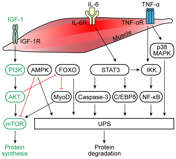
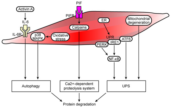
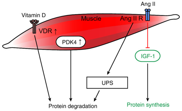
Muscle atrophy is a severe clinical problem involving the loss of muscle mass and strength that frequently accompanies the development of numerous types of cancer, including pancreatic, lung and gastric cancers. Cancer cachexia is a multifactorial syndrome characterized by a continuous decline in skeletal muscle mass that cannot be reversed by conventional nutritional therapy. The pathophysiological characteristic of cancer cachexia is a negative protein and energy balance caused by a combination of factors, including reduced food intake and metabolic abnormalities. Numerous necessary cellular processes are disrupted by the presence of abnormal metabolites, which mediate several intracellular signaling pathways and result in the net loss of cytoplasm and organelles in atrophic skeletal muscle during various states of cancer cachexia. Currently, the clinical morbidity and mortality rates of patients with cancer cachexia are high. Once a patient enters the cachexia phase, the consequences are difficult to reverse and the treatment methods for cancer cachexia are very limited. The present review aimed to summarize the recent discoveries regarding the pathogenesis of cancer cachexia‑induced muscle atrophy and provided novel ideas for the comprehensive treatment to improve the prognosis of affected patients.


Figures

Figure 1.
Figure 1.
Molecules and signaling pathways involved in muscle protein synthesis and degradation. Under physiological conditions, IGF-1 activates AKT through a PI3K-dependent process, leading to the activation of mTOR and thus resulting in the increased proliferation of muscle cells and increased protein synthesis in muscle cells. IL-6 and TNF-α are considered to be the main mediators of the inflammation in muscle atrophy caused by cancer cachexia. The binding of IL-6 to its receptor induces AMPK and STAT3 expression. STAT3 induces the activation of the IKK/NF-κB pathway and caspase-3 and C/EBPδ expression, which activates the UPS, causing muscle protein degradation. AMPK is a downstream target of IL-6 signaling, which inhibits the mTOR cascade, and activates the UPS. Physiologically, AKT inhibits FOXO, which promotes protein synthesis. In cancer cachexia, FOXO inhibits MyoD and activates the UPS, thereby promoting protein degradation. Moreover, the activation of NF-κB due to the degradation of the IκB inhibitor by IKK is TNF-α-dependent. The NF-κB pathway can further activate the UPS to cause muscle protein degradation. TNF-α can also induce the p38 MAPK pathway, which activates the UPS. IGF-1, insulin-like growth factor 1; AMPK, AMP-activated protein kinase; IKK, IκB kinase; C/EBPδ, CCAAT/enhancer-binding protein δ; UPS, ubiquitin-proteasome system; MyoD, myoblast determination protein 1; R, receptor.
Figure 2.
Figure 2.
Cell autophagy/lysosomal and Ca2+-dependent protein degradation pathways, ER stress and mitochondrial dysfunction are involved in muscle protein degradation in cancer cachexia. In cancer cachexia, increased activin A expression activates the IL-6 signaling pathway, and oxidative stress induces activation of the p38 MAPK signaling pathway, resulting in autophagy and protein degradation. The overexpression of Ca2+-dependent proteases (calpains) activates the Ca2+-dependent proteolysis system, resulting in increased protein degradation. In addition, the ER manages such stress by initiating the UPR, which is controlled by three transmembrane proteins, namely, PERK, IRE1 and ATF6. Optimal activation of NF-κB during ER stress requires inputs from both IRE1 and PERK activities, and ATF6 may interact with protein degradation pathways, such as the UPS. Moreover, local mitochondrial degeneration in the muscle activates the UPS, which results in protein degradation. R, receptor; PERK, protein kinase-like ER eukaryotic translation initiation factor 2α kinase; ER, endoplasmic reticulum; UPR, unfolded protein response; IRE1, inositol-requiring protein; ATF6, activating transcription factor 6; UPS, ubiquitin-proteasome system.
Figure 3.
Figure 3.
Receptors and substances involved in metabolism that affect muscle protein synthesis and degradation in cancer cachexia. In cancer cachexia, the overexpression of the VDR impairs muscle regeneration and causes protein degradation. Increased PDK4 expression directly promotes cancer cachexia-related changes in muscle metabolism and skeletal muscle atrophy. Furthermore, Ang II activates the UPS to induce protein degradation and inhibits the IGF-1 signaling pathway, thereby interfering with protein synthesis. VDR, vitamin D receptor; Ang II, angiotensin II; R, receptor; UPS, ubiquitin-proteasome system; IGF-1, insulin-like growth factor-1; PDK4, pyruvate dehydrogenase kinase 4.

References

    1. Fearon K, Strasser F, Anker SD, Bosaeus I, Bruera E, Fainsinger RL, Jatoi A, Loprinzi C, MacDonald N, Mantovani G, et al. Definition and classification of cancer cachexia: An international consensus. Lancet Oncol. 2011;12:489–495. doi: 10.1016/S1470-2045(10)70218-7. - DOI - PubMed
    1. Blum D, Stene GB, Solheim TS, Fayers P, Hjermstad MJ, Baracos VE, Fearon K, Strasser F, Kaasa S; Euro-Impact. Validation of the consensus-definition for cancer cachexia and evaluation of a classification model-a study based on data from an international multicentre project (EPCRC-CSA) Ann Oncol. 2014;25:1635–1642. doi: 10.1093/annonc/mdu086. - DOI - PubMed
    1. Baumgartner RN, Koehler KM, Gallagher D, Romero L, Heymsfield SB, Ross RR, Garry PJ, Lindeman RD. Epidemiology of sarcopenia among the elderly in New Mexico. Am J Epidemiol. 1998;147:755–763. doi: 10.1093/oxfordjournals.aje.a009520. - DOI - PubMed
    1. Emery PW, Edwards RH, Rennie MJ, Souhami RL, Halliday D. Protein synthesis in muscle measured in vivo in cachectic patients with cancer. Br Med J (Clin Res Ed) 1984;289:584–586. doi: 10.1136/bmj.289.6445.584. - DOI - PMC - PubMed
    1. Warnold I, Lundholm K, Schersten T. Energy balance and body composition in cancer patients. Cancer Res. 1978;38:1801–1807. - PubMed