. 2021 Feb 11;155(3):354-363.
doi: 10.1093/ajcp/aqaa230.

A Descriptive and Quantitative Immunohistochemical Study Demonstrating a Spectrum of Platelet Recruitment Patterns Across Pulmonary Infections Including COVID-19

Affiliations

A Descriptive and Quantitative Immunohistochemical Study Demonstrating a Spectrum of Platelet Recruitment Patterns Across Pulmonary Infections Including COVID-19

Phillip D McMullen et al. Am J Clin Pathol. .

Abstract

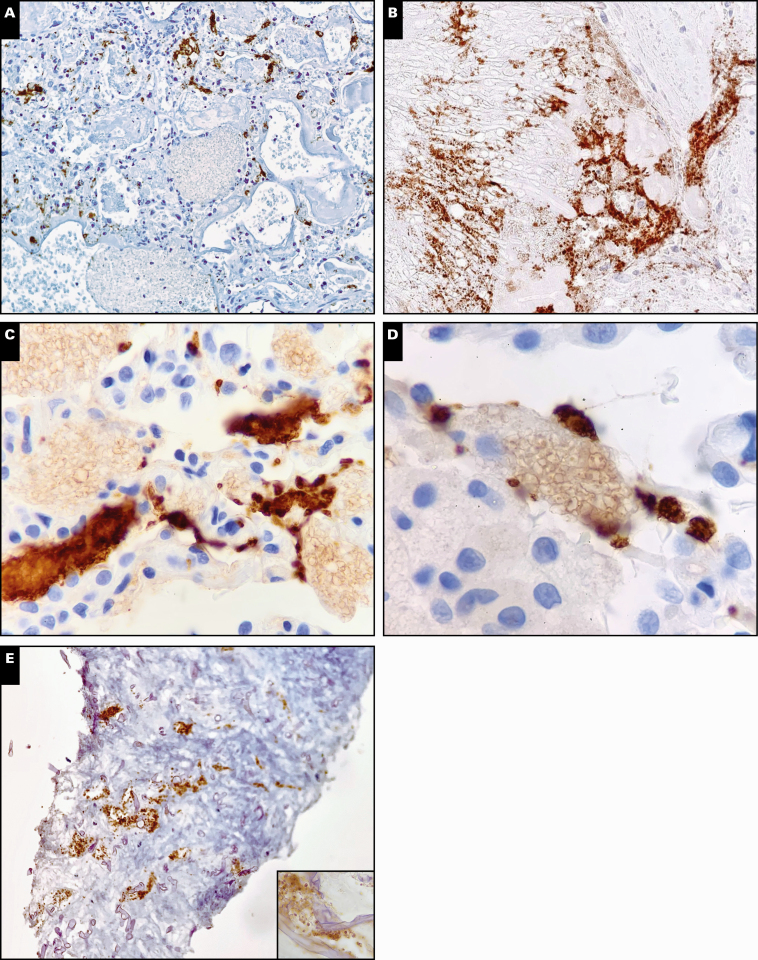
Objectives: Pulmonary platelet deposition and microangiopathy are increasingly recognized components of coronavirus disease 2019 (COVID-19) infection. Thrombosis is a known component of sepsis and disseminated intravascular coagulation. We sought to compare the level of platelet deposition in the pulmonary vasculature in cases of confirmed COVID-19 infection to other lung injuries and infections.

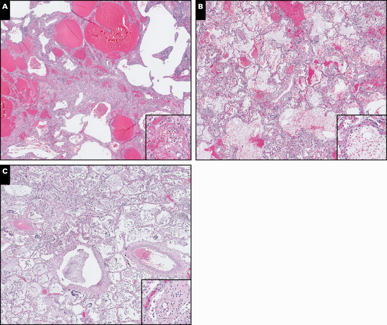
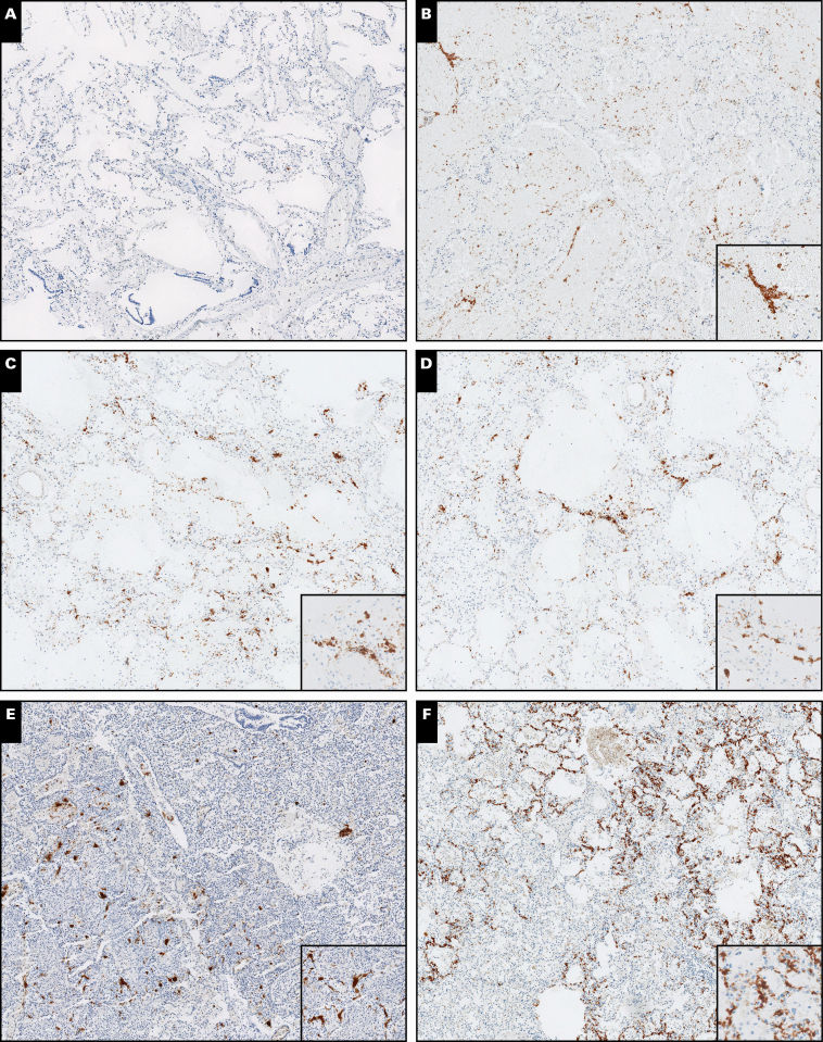
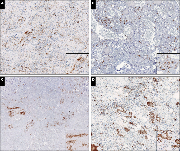
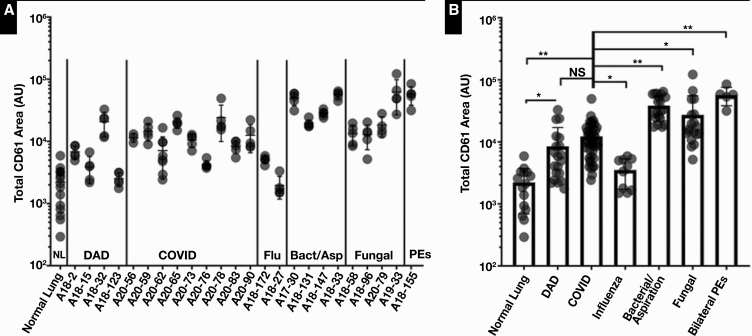
Methods: Immunohistochemistry was performed on 27 autopsy cases and 2 surgical pathology cases targeting CD61. Multiple cases of normal lung, diffuse alveolar damage, COVID-19, influenza, and bacterial and fungal infections, as well as one case of pulmonary emboli, were included. The levels of CD61 staining were compared quantitatively in the autopsy cases, and patterns of staining were described.

Results: Nearly all specimens exhibited an increase in CD61 staining relative to control lung tissue. The area of CD61 staining in COVID-19 infection was higher than influenza but still comparable to many other infectious diseases. Cases of aspiration pneumonia, Staphylococcus aureus infection, and blastomycosis exhibited the highest levels of CD61 staining.

Conclusions: Platelet deposition is a phenomenon common to many pulmonary insults. A spectrum of staining patterns was observed, suggestive of pathogen-specific mechanisms of platelet deposition. Further study into the mechanisms driving platelet deposition in pulmonary injuries and infections is warranted.

Keywords: CD61; COVID-19; Coronavirus; Diffuse alveolar damage; Platelets; Pulmonary intravascular coagulopathy.


Figures

Image 1
Image 1
A-C, Representative H&E-stained slides of coronavirus disease 2019 infections demonstrating diffuse alveolar damage and hemorrhagic changes (H&E, ×5; inset, ×20).
Image 2
Image 2
Representative scanned images of CD61-stained tissue from autopsy specimens. A, Control lung. B, Diffuse alveolar damage (noninfectious). C, Coronavirus disease 2019. D, Influenza. E, Pseudomonas pneumonia. F, Methicillin-sensitive Staphylococcus aureus pneumonia. All images were taken at ×5 magnification (insets, approximately ×20).
Image 3
Image 3
Representative scanned images of CD61-stained tissue from autopsy specimens. A, Aspiration pneumonia. B, Candidiasis. C, Aspergillosis. D, Multiple pulmonary emboli. All images were taken at ×5 magnification (insets, approximately ×20).
Figure 1
Figure 1
Results of quantitative immunohistochemistry analysis of CD61-stained tissue sections across all cases (A) and groups (B). Points represent individual measurements obtained from still images, and error bars represent standard deviation. Statistical analysis was performed using a one-way analysis of variance (P < .0001) for comparison of all cases and unpaired Student t tests for individual comparisons. Statistical significance is abbreviated as follows: NS, not significant, *P < .01, **P < .0001. AU, arbitrary units; Bact/Asp, bacterial/aspiration; DAD, noninfectious causes of diffuse alveolar damage; Flu, influenza; NL, normal lung; PEs, pulmonary emboli.
Image 4
Image 4
Examples of pathogen-specific staining patterns identified with CD61 immunostaining. A, Discrete staining of capillaries adjacent to yeast colony, with faint surface staining of yeast forms adjacent to platelets (×10). B, Large patch of positive staining likely representing angioinvasion in aspergillosis, with direct platelet-hyphal interactions observed (×20). C, D, Platelet aggregates adjacent to fungal colonies with surface staining of yeasts in pneumocystis infection (×40). E, Staining of vessels at the periphery of invasive zygomycosis and evidence of direct fungal-hyphae interactions (×20; inset, ×40).

Comment in

  • Platelet Recruitment in COVID-19.
    Cardillo A, Refaai M. Cardillo A, et al. Am J Clin Pathol. 2022 Jan 6;157(1):153. doi: 10.1093/ajcp/aqab102. Am J Clin Pathol. 2022. PMID: 34463341 Free PMC article. No abstract available.

References

    1. Llitjos J-F, Leclerc M, Chochois C, et al. High incidence of venous thromboembolic events in anticoagulated severe COVID-19 patients. J Thromb Haemost. 2020;18:1743-1746. - PMC - PubMed
    1. Bikdeli B, Madhavan MV, Jimenez D, et al. COVID-19 and thrombotic or thromboembolic disease: implications for prevention, antithrombotic therapy, and follow-up. J Am Coll Cardiol. 2020;75:2950-2973. - PMC - PubMed
    1. Magro C, Mulvey JJ, Berlin D, et al. Complement associated microvascular injury and thrombosis in the pathogenesis of severe COVID-19 infection: a report of five cases. Transl Res. 2020;220:1-13. - PMC - PubMed
    1. Klok FA, Kruip MJHA, van der Meer NJM, et al. Incidence of thrombotic complications in critically ill ICU patients with COVID-19. Thromb Res. 2020;191:145-147. - PMC - PubMed
    1. Escher R, Breakey N, Lämmle B. Severe COVID-19 infection associated with endothelial activation. Thromb Res. 2020;190:62. - PMC - PubMed