Review
. 2020 Nov 11;11(1):116.
doi: 10.1186/s13244-020-00916-0.

Radiological manifestations of thoracic hydatid cysts: pulmonary and extrapulmonary findings

Affiliations
Review

Radiological manifestations of thoracic hydatid cysts: pulmonary and extrapulmonary findings

Gamze Durhan et al. Insights Imaging. .

Abstract

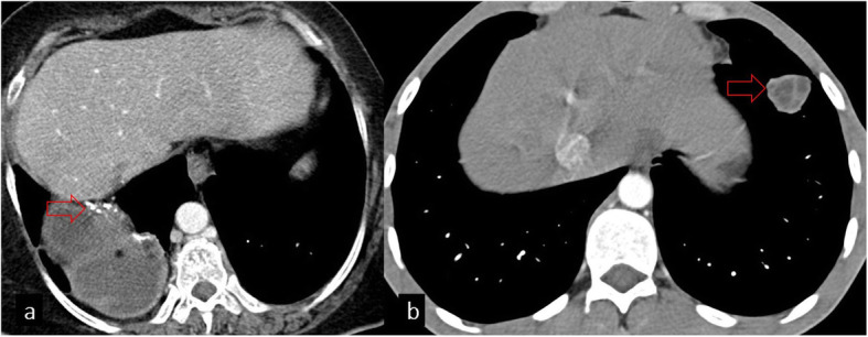
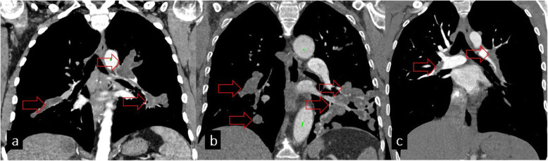
Hydatid cyst caused by the larval form of Echinococcus is a worldwide zoonosis. The lungs and liver are the most common sites involved. While the lung parenchyma is the most common site within the thorax, it may develop in any extrapulmonary region including the pleural cavity, fissures, mediastinum, heart, vascular structures, chest wall, and diaphragm. Imaging plays a pivotal role not only in the diagnosis of hydatid cyst, but also in the visualization of the extent of involvement and complications. The aim of this pictorial review was to comprehensively describe the imaging findings of thoracic hydatid cyst including pulmonary and very unusual extrapulmonary involvements. An outline is also given for the findings of complications and differential diagnosis of thoracic hydatid cyst.

Keywords: Extrapulmonary; Pulmonary; Thoracic hydatid cyst.


Conflict of interest statement

The authors declare that they have no competing interests.

Figures

Fig. 1
Fig. 1
Hydatid cyst has a non-specific appearance on chest radiography
Fig. 2
Fig. 2
a Coronal chest CT image in the mediastinal window shows a giant (> 10 cm) uncomplicated hydatid cyst (red arrow). Measurement of HU values on CT shows low density (mean HU ± SD = − 1 ± 10) consistent with simple fluid. b On axial CT image, bilateral hydatid cysts are seen (red arrows)
Fig. 3
Fig. 3
Radiological findings of pulmonary hydatid cysts
Fig. 4
Fig. 4
a Calcification in the wall of the hydatid cyst is seen on the axial CT image (red arrow). b Daughter cysts in the hydatid cyst (red arrow)
Fig. 5
Fig. 5
Imaging features of contained rupture. Axial CT images show the crescent sign (a, red arrow), inverse crescent sign (b, red arrow), and air bubble sign (c, red arrow)
Fig. 6
Fig. 6
Complete rupture signs. Axial CT images of different patients on the lung window (ae). a Cumbo sign (red arrow). b Mass within cavity sign (red arrow). c Waterlily sign (red arrow). d Whirl sign (red arrow) e. Empty cyst sign (red arrow)
Fig. 7
Fig. 7
Imaging features of hydatid cyst rupture directly to the lung parenchyma. Axial CT images of different patients showing centrilobular nodular opacities around the hydatic cyst (a, red arrow), tree-in-bud pattern around the HC (b, red arrow), and consolidation around the HC (c, red arrow)
Fig. 8
Fig. 8
Axial CT images in the lung (a, b) and mediastinal (c, d) windows show associated findings of HC. a Atelectasis. b Bronchiectasis. c Mediastinal lymphadenopathy seen especially in hydatic cyst with superinfection. d Pleural effusion
Fig. 9
Fig. 9
Steady-state free precession (SSFP) gradient echo magnetic resonance image shows a hyperintense hydatid cyst in the left pulmonary artery (a), contrast-enhanced T1W image shows hypointensity (b), and gradient TRUFI demonstrates hyperintensity in hydatid cysts (c)
Fig. 10.
Fig. 10.
Axial CT image shows an opacification with smooth contours, which is a pathologically proven bronchogenic cyst (red arrow)
Fig. 11
Fig. 11
Bone hydatid cysts and differential diagnosis. a Axial chest CT image shows an HC located anteriorly adjacent to the sternum causing sternal cortical erosion (red arrow) b Axial CT image in the bone window shows the destruction of the vertebra and adjacent costa (red arrows) secondary to HC. c Costa destruction secondary to lung cancer is seen (red arrow)
Fig. 12
Fig. 12
Hydatid cysts in thoracic soft tissues and differential diagnosis. a Axial CT image shows left paraspinal muscle involvement of HC (red arrow). b Pectoral hematoma as a differential diagnosis of HC seen on CT image (red arrow). c HC with daughter cysts located in the right axilla seen on the axial CT image (red arrow). d Necrotic lymphadenopathy secondary to lung cancer is seen in the right axilla
Fig. 13
Fig. 13
a Hydatid cysts of the pleura and fissure. b Right pleural cystic lesion with peripheral contrast enhancement that belongs to empyema
Fig. 14
Fig. 14
Chest CT images on the coronal plane show transdiaphragmatic migration of hydatid cysts in different patients (a, b, red arrows). Diaphragmatic discontinuation can be observed
Fig. 15
Fig. 15
a Axial CT image shows a cystic lesion in the mediastinum, adjacent to the left ventricle, pathologically proven as a hydatid cyst (red arrow). b, c, d Differential diagnosis of mediastinal hydatid cysts: thymoma (b), pericardial cyst (c), and teratoma (d)
Fig. 16
Fig. 16
Cardiac involvement of hydatid cyst. a Hydatid cyst with calcification in the left ventricle is seen on the short-axis CT image (red arrow). b Hydatid cyst in the left ventricle is demonstrated in another patient (red arrow)
Fig. 17
Fig. 17
a Hydatid cysts of pulmonary arteries seen on the coronal chest CT image. b Sarcoma of the pulmonary artery shows vascular distension and fills the lumen like hydatid cysts of the pulmonary arteries. c Pulmonary thromboembolism as another differential diagnosis of hydatid cyst seen on the coronal chest CT image
Fig. 18
Fig. 18
Axial chest CT image in the mediastinal window shows pseudoaneurysm formation and wall erosion in ascending aorta (red arrow)

References

    1. Neva FA (1994) BHIhIBpN, Conn. Appleton&Lange 128-133
    1. Pedrosa I, Saiz A, Arrazola J, Ferreiros J, Pedrosa CS. Hydatid disease: radiologic and pathologic features and complications. Radiographics. 2000;20:795–817. doi: 10.1148/radiographics.20.3.g00ma06795. - DOI - PubMed
    1. Haliloglu M, Saatci I, Akhan O, Ozmen MN, Besim A. Spectrum of imaging findings in pediatric hydatid disease. AJR Am J Roentgenol. 1997;169:1627–1631. doi: 10.2214/ajr.169.6.9393178. - DOI - PubMed
    1. Beggs I. The radiology of hydatid disease. AJR Am J Roentgenol. 1985;145:639–648. doi: 10.2214/ajr.145.3.639. - DOI - PubMed
    1. Mehta P, Prakash M, Khandelwal N. Radiological manifestations of hydatid disease and its complications. Trop Parasitol. 2016;6:103–112. doi: 10.4103/2229-5070.190812. - DOI - PMC - PubMed

LinkOut - more resources