. 2020 Nov 11;15(11):e0240347.
doi: 10.1371/journal.pone.0240347. eCollection 2020.

Organizing pneumonia of COVID-19: Time-dependent evolution and outcome in CT findings

Affiliations

Organizing pneumonia of COVID-19: Time-dependent evolution and outcome in CT findings

Yan Wang et al. PLoS One. .

Abstract

Background: As a pandemic, a most-common pattern resembled organizing pneumonia (OP) has been identified by CT findings in novel coronavirus disease (COVID-19). We aimed to delineate the evolution of CT findings and outcome in OP of COVID-19.

Materials and methods: 106 COVID-19 patients with OP based on CT findings were retrospectively included and categorized into non-severe (mild/common) and severe (severe/critical) groups. CT features including lobar distribution, presence of ground glass opacities (GGO), consolidation, linear opacities and total severity CT score were evaluated at three time intervals from symptom-onset to CT scan (day 0-7, day 8-14, day > 14). Discharge or adverse outcome (admission to ICU or death), and pulmonary sequelae (complete absorption or lesion residuals) on CT after discharge were analyzed based on the CT features at different time interval.

Results: 79 (74.5%) patients were non-severe and 103 (97.2%) were discharged at median day 25 (range, day 8-50) after symptom-onset. Of 67 patients with revisit CT at 2-4 weeks after discharge, 20 (29.9%) had complete absorption of lesions at median day 38 (range, day 30-53) after symptom-onset. Significant differences between complete absorption and residuals groups were found in percentages of consolidation (1.5% vs. 13.8%, P = 0.010), number of involved lobe > 3 (40.0% vs. 72.5%, P = 0.030), CT score > 4 (20.0% vs. 65.0%, P = 0.010) at day 8-14.

Conclusion: Most OP cases had good prognosis. Approximately one-third of cases had complete absorption of lesions during 1-2 months after symptom-onset while those with increased frequency of consolidation, number of involved lobe > 3, and CT score > 4 at week 2 after symptom-onset may indicate lesion residuals on CT.


Conflict of interest statement

The authors have declared that no competing interests exist.

Figures

Fig 1
Fig 1. Patients recruitment flowchart.
COVID-19 = coronavirus disease 2019, ICU = intensive care unit, OP = organizing pneumonia.
Fig 2
Fig 2. The evolution of CT findings across the three time groups (day 0–7, day 8–14, day >14) in COVID-19 patients with organizing pneumonia pattern.
GGO = ground glass opacity; with three signs = GGO, consolidation and linear opacity.
Fig 3
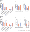
Fig 3. Comparisons of CT findings between patients with complete absorption and residuals after discharge.
GGO = ground glass opacity; with three signs = GGO, consolidation and linear opacity. *, P< 0.05 indicated significant differences.
Fig 4
Fig 4. A 66-year-old man who had been to Wuhan had fever for 4 days and discharged at day 28 after symptom onset.
(A) CT obtained on admission at day 4 after symptom onset shows multiple subpleural consolidation. (B) CT on day 11 shows progression with increased number and size of lung lesions. (C, E) CT on day 27 shows absorption with decreased density of the lesions. (D) CT on day 41 shows almost complete absorption of pulmonary lesions.
Fig 5
Fig 5. A 55-year-old man who had fever for 6 days and discharged at day 31 after symptom onset.
(A) CT obtained on admission at day 6 after symptom onset shows focal GGO in bilateral lower lobe. (B) CT on day 10 shows progression with increased size and density of lung lesions. (C) CT on day 18 shows progression with increased density of the lesions. (D) CT on day 30 shows patchy consolidation and linear opacities of pulmonary lesions.

References

    1. Zhu N, Zhang D, Wang W, Li X, Yang B, Song J. et al. A Novel Coronavirus from Patients with Pneumonia in China, 2019. N. Engl. J. Med. 2020;382(8):727–733. 10.1056/NEJMoa2001017 - DOI - PMC - PubMed
    1. Huang C, Wang Y, Li X, Ren L, Zhao J, Hu Y. et al. Clinical features of patients infected with 2019 novel coronavirus in Wuhan, China . Lancet. 2020. Feb 15; 395(10223):497–506. 10.1016/S0140-6736(20)30183-5 - DOI - PMC - PubMed
    1. Guan W, Ni Z, Hu Y, Liang W, Ou C, He J. et al. Clinical characteristics of 2019 novel coronavirus infection in China. N Engl J Med. 2020. Apr 30;382(18):1708–1720. 2020. 10.1056/NEJMoa2002032 - DOI - PMC - PubMed
    1. WHO. Coronavirus disease 2019. 2020. https://www.who.int/emergencies/diseases/novel-coronavirus-2019/situatio.... Accessed April 29, 2020.
    1. Kanne JP, Little BP, Chung JH, Elicker BM, Ketai LH. Essentials for Radiologists on COVID-19: An Update—Radiology Scientific Expert Panel. Radiology. 2020. Feb 27:200527 10.1148/radiol.2020200527 - DOI - PMC - PubMed

MeSH terms

Substances