Review
. 2021 Jan 1;320(1):C92-C105.
doi: 10.1152/ajpcell.00355.2020. Epub 2020 Nov 11.

Vessel-on-a-chip models for studying microvascular physiology, transport, and function in vitro

Affiliations
Review

Vessel-on-a-chip models for studying microvascular physiology, transport, and function in vitro

Savannah R Moses et al. Am J Physiol Cell Physiol. .

Abstract

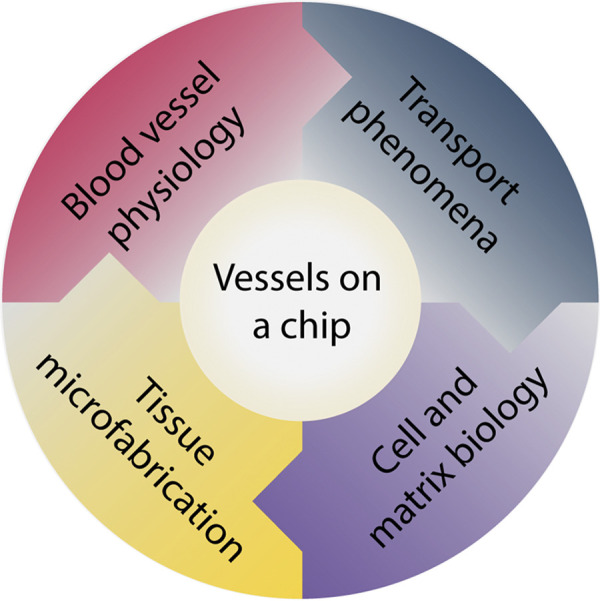
To understand how the microvasculature grows and remodels, researchers require reproducible systems that emulate the function of living tissue. Innovative contributions toward fulfilling this important need have been made by engineered microvessels assembled in vitro with microfabrication techniques. Microfabricated vessels, commonly referred to as "vessels-on-a-chip," are from a class of cell culture technologies that uniquely integrate microscale flow phenomena, tissue-level biomolecular transport, cell-cell interactions, and proper three-dimensional (3-D) extracellular matrix environments under well-defined culture conditions. Here, we discuss the enabling attributes of microfabricated vessels that make these models more physiological compared with established cell culture techniques and the potential of these models for advancing microvascular research. This review highlights the key features of microvascular transport and physiology, critically discusses the strengths and limitations of different microfabrication strategies for studying the microvasculature, and provides a perspective on current challenges and future opportunities for vessel-on-a-chip models.

Keywords: angiogenesis; cellular microenvironment; microfluidics; tissue microfabrication; vascular remodeling.


Conflict of interest statement

No conflicts of interest, financial or otherwise, are declared by the authors.

Figures

Figure 1.
Figure 1.
Integrative vessel-on-a-chip models. These engineered models are due to the convergence of principles from 1) blood vessel physiology, 2) transport phenomena, 3) cell and matrix biology, and 4) tissue microfabrication.
Figure 2.
Figure 2.
Mass transport in the arterial microcirculation. A: the largest vessels in the arterial microcirculation are arterioles or “resistance vessels,” which supply blood to capillaries via convection (Pe > 1). A circumferential layer of smooth muscle cells enables precise mechanical control over blood flow and subsequent regional perfusion. In normal physiological environments, surrounding tissue and ECM is normoxic [partial pressure of oxygen (PO2) ∼ 5%] (38). B: capillaries are the smallest vessels in the microcirculation and the main site for gas and nutrient exchange. A thin endothelial vessel wall enables passive diffusion (Pe < 1). The surrounding ECM is normoxic, with a higher PO2 nearest the capillary as O2 is radially dispersed. C: tissue hypoxia (PO2 < 5%) drives vessel angiogenesis in postcapillary venules. Increased vessel permeability causes leakage of plasma proteins (green) into surrounding interstitial fluid. Coupled with infiltration of endothelial cells (pink), these components assemble to form new vascular sprouts. (Drawing is not to scale.)
Figure 3.
Figure 3.
On-chip devices used to mimic various tissue/vascular functional barriers. A: blood brain barrier microfluidic model used to study nutrient exchange/waste removal in order to develop new drug delivery therapies. B: alveolar-capillary interface microfluidic model used to study pulmonary physiology in patients with thrombotic diseases. C: kidney-on-a-chip model utilized to study blood filtration and waste removal. D: human placenta-on-a-chip model designed to examine the effects of nanoparticles transferred from the mother to the fetus via the placenta. In this model, human placental trophoblast BeWo cells are cocultured with human umbilical vein endothelial cells (HUVECs). [Fig. 3A reproduced from Moya et al. (175), Fig. 3B from Jain et al. (174), Fig. 3C from Rayner et al. (176), and Fig. 3D from Yin et al. (177), with permission.]

References

    1. Adams RH, Alitalo K. Molecular regulation of angiogenesis and lymphangiogenesis. Nat Rev Mol Cell Biol 8: 464–478, 2007. doi:10.1038/nrm2183. - DOI - PubMed
    1. Carmeliet P, Jain RK. Molecular mechanisms and clinical applications of angiogenesis. Nature 473: 298–307, 2011. doi:10.1038/nature10144. - DOI - PMC - PubMed
    1. Goldman D. Theoretical models of microvascular oxygen transport to tissue. Microcirculation 15: 795–811, 2008. doi:10.1080/10739680801938289. - DOI - PMC - PubMed
    1. Popel AS. Theory of oxygen transport to tissue. Crit Rev Biomed Eng 17: 257–321, 1989. - PMC - PubMed
    1. Fleetwood IG, Steinberg GK. Arteriovenous malformations. Lancet 359: 863–873, 2002. doi:10.1016/S0140-6736(02)07946-1. - DOI - PubMed

Publication types

LinkOut - more resources