. 2020 Nov 10;33(6):108368.
doi: 10.1016/j.celrep.2020.108368.

Intramacrophage ROS Primes the Innate Immune System via JAK/STAT and Toll Activation

Affiliations

Intramacrophage ROS Primes the Innate Immune System via JAK/STAT and Toll Activation

Sveta Chakrabarti et al. Cell Rep. .

Abstract

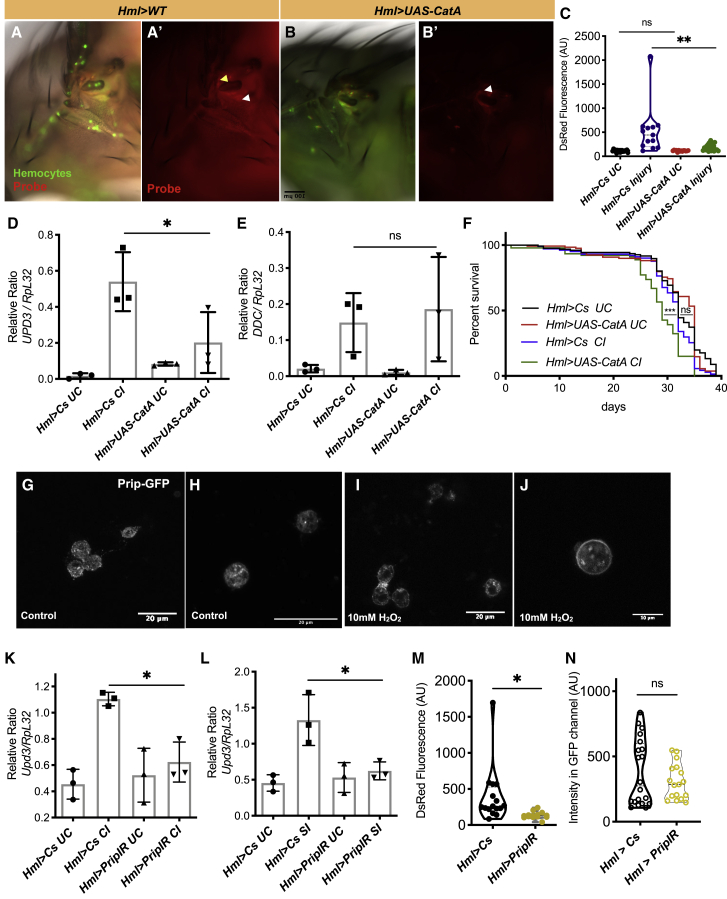
Tissue injury is one of the most severe environmental perturbations for a living organism. When damage occurs in adult Drosophila, there is a local response of the injured tissue and a coordinated action across different tissues to help the organism overcome the deleterious effect of an injury. We show a change in the transcriptome of hemocytes at the site of tissue injury, with pronounced activation of the Toll signaling pathway. We find that induction of the cytokine upd-3 and Toll receptor activation occur in response to injury alone, in the absence of a pathogen. Intracellular accumulation of hydrogen peroxide in hemocytes is essential for upd-3 induction and is facilitated by the diffusion of hydrogen peroxide through a channel protein Prip. Importantly, hemocyte activation and production of reactive oxygen species (ROS) at the site of a sterile injury provide protection to flies on subsequent infection, demonstrating training of the innate immune system.

Keywords: Drosophila; JAK/STAT signaling; Toll pathway; hemocyte; injury; reactive oxygen species; trained immunity; upd-3.


Conflict of interest statement

Declaration of Interests The authors declare no competing interests.

Figures

None
Graphical abstract
Figure 1
Figure 1
Defense Pathway Toll and Oxidative Stress Responses Are Induced upon Wounding in Hemocytes (A) A volcano plot showing genes significantly altered in hemocytes 1 h after injury. Log2 fold change values are plotted on the horizontal axis and −log10 of the p value on the vertical axis. Statistically significant regulated genes (p < 0.01) are shown in red, where GO categories are indicated (defense response, purple; membrane proteins, cyan; neuronal, yellow; redox, green; and phagocytosis, blue). (B) Validation of maximally differentially expressed genes from the RNA-seq data by qRT-PCR experiments in wild-type (w1118) hemocytes following injury. CecC, DptA, AttB, and CG16772 were found to be significantly upregulated by qRT-PCR. ∗∗∗p < 0.0001; p < 0.05; ns, non-significant. Female flies were used for experiments. (C) qRT-PCR of the AMPs CecC and AttB expression in axenic flies in comparison with conventionally reared w1118 flies. UC, unchallenged; CI, clean injury. (D) Differentially regulated genes from the RNA-seq experiment according to their GO categories. Gene names, functions, fold change expression, and q-values are denoted for different GO categories, including the defense response. (E) Duox, NOX, and IRC gene expression (top) and upd-1, upd-2, and upd-3 gene expression (bottom) from the RNA-seq data in wild-type (w1118) hemocytes upon injury. IRC and upd-3 were found to be significantly upregulated. p < 0.05, as determined by Student’s t test. Counts were normalized and extracted using the software featureCounts (PMID: 24227677). ± SD are shown in (B) and (C).
Figure 2
Figure 2
Production of Hydrogen Peroxide from a Wound Activates Hemocytes and Increases Expression of upd-3 (A and B) Representative images of a TCFB probe (in red) before and after injury to the thorax in hemocytes using Hml:UAS-GFP (green) female flies. Hydrogen peroxide accumulates at the site of injury as well as in hemocytes located at that site. The TCFB probe (in red) is shown for the site of injury. (C and D) Probe fluorescence in Hml>UAS-DuoxIR flies and Hml>UAS-IRC flies within hemocytes and around the wound in the cuticle. White arrows mark cuticular ROS in (B′), (C′), and (D′), while yellow arrows mark hemocyte accumulated probe in (B′) and (D′). (E and F) Quantification of the increase in fluorescence using arbitrary units in ∼30 flies per condition and per genotype. Decreased probe fluorescence in Hml>UAS-DuoxIR (red) and Hml>UAS-GFP, UAS-IRC flies (green) was observed. (G and H) Quantification of the GFP fluorescence using arbitrary units in ∼20 flies per condition around the site of injury in Hml>Cs and Hml>UAS-DuoxIR. The number of hemocytes at the site of the injury was not changed upon Duox knockdown from hemocytes. (I) Flies with reduced ROS burst after an injury (i.e., Hml>UAS-DuoxIR and Hml>UAS-IRC) show increased susceptibility to injury. Flies per genotype from at least five independent experiments were Hml>UAS-DuoxIR (n = 93), Hml>UAS-IRC (n = 88), and wild type (Hml>Cs, n = 138). (J) Expression of upd-3 in hemocytes in Hml>UAS-DuoxIR and hmlΔGAL4>UAS-IRC adult flies. (K) Ectopic overexpression of Duox in adult hemocytes is sufficient to stimulate the expression of upd-3 in hemocytes without any injury. For (E)–(K) mean values of at least three experiments (with 30–40 flies each) ± SD are shown. ∗∗p < 0.01; p < 0.05; ∗∗∗p < 0.001; ns, non-significant.
Figure 3
Figure 3
Intracellular ROS Is Required for Activation of Macrophages and Survival of Flies following Injury (A and B) Overexpression of CatA leads to the reduction of ROS in the cytoplasm of hemocytes. Quantification done similar to Figure 2E. White arrows mark cuticular ROS on (A′), (B′), and (D′), while the yellow arrow marks hemocyte accumulated probe in (A′). (C) Decreased probe fluorescence in Hml>UAS-CatA flies upon injury as compared to their wild-type counterparts. (D and E) Expression of upd-3 with and without injury and ddc expression after injury in hemocytes as compared to their wild-type counterparts (Hml>UAS-CatA versus Hml>CS). (F) Reduced intracellular ROS accumulation in hemocytes after an injury in Hml>UAS-CatA leads to increased susceptibility to injury. Hml>UAS-CatA (n = 70 [UC]; n = 76 [CI] and wild-type [Hml>Cs, n = 80 (UC); n = 70 (CI)] adult flies. UC, unchallenged; CI, clean injury. (G–J) Prip channel localization in hemocytes. Panels indicate the distribution of Prip localization in hemocytes in control media (top) and media containing 10 mM H2O2 (I and J). Membrane localization of Prip in an enlarged image is shown (J). (K) Quantification of the increase in fluorescence of the TCFB probe after an injury to the thorax in hemocytes in prip knockdown flies. (L) Quantification of the increase in GFP fluorescence around the wounded area in Hml:UAS-GFP animals and prip knockdown flies in at least 20 flies per condition. (M) qRT-PCR in wild-type flies and Hml>UAS-Prip-IR. (N) qRT-PCR in wild-type flies and Hml>UAS-Prip-IR. upd-3 is not induced after septic injury in knocked down flies. Similar results were obtained with a second RNAi line against Prip-IR line (BDSC_50695). ∗∗p < 0.01; p < 0.05; ns, non-significant. ± SD are shown in (D), (E), (M), and (N).
Figure 4
Figure 4
The Src42A/Shark/Draper Pathway Regulates upd-3 Expression in Hemocytes (A, E, and G) Lifespan of drprΔ5 flies and susceptibility to injury as compared to the wild type (w1118). Flies per genotype are pooled from at least three independent experiments. Log-rank test was used for comparing wild-type (w1118; CI, n = 112; SI, n = 63) and drprΔ5 (CI, n = 129; SI, n = 61) flies; Hml>UAS-drprIR (n = 81) and Hml>UAS-Src42AIR (n = 62) adult flies as compared to wild type (Hml>Cs, n = 173). UC, unchallenged; CI, clean injury; SI, septic injury. (B and C) qRT-PCR of upd-3 in drprΔ5 mutant flies 1 h after injury (B) and septic injury (C). (D, F, and H) Hemocyte knockdown of drprIR (D), src42AIR (F), and shark1 heterozygotes (H) shows a decreased expression of upd-3 expression 1 h after injury (±SD is shown). ∗∗∗p < 0.0001; ∗∗p < 0.01; p < 0.05; ns, non-significant.
Figure 5
Figure 5
Injury Protects Flies from a Subsequent Systemic Infection (A) Experimental setup for determining immune training after an injury to a pathogen systemic infection. Flies were either injured or left undisturbed prior to infection with E. faecalis. (B) Survival of flies following infection with E. faecalis as indicated in (A) (w1118; UC, n = 58; CI, n = 44). (C–F) qRT-PCR of the AMPs CecC and drosomycin (drs) expression in hemocytes and fat body of naive versus injured flies w1118 flies that were subsequently infected with E. faecalis for 20 h. (G) Survival of drprΔ5 mutant flies with E. faecalis as indicated in (A). (H) qRT-PCR in wild-type and drprΔ5 mutant flies in hemocytes of naive versus injured flies that were subsequently infected with E. faecalis for 20 h. The loss of response to the ROS signal upon wounding in leads to an attenuated hemocyte AMP activation to infection with E. faecalis. UC, unchallenged; CI: clean injury; Ef, E. faecalis. For (C)–(F) and (H), data represent ±SD. p < 0.05; ns, non-significant.
Figure 6
Figure 6
Features of a Response to Systemic Injury in Flies Following injury, hemocytes respond to the damage signal H2O2, which is produced by the NADPH oxidase Duox, through the kinase Src42A and its downstream targets Shark and Draper. A channel protein, Prip, helps increase intracellular ROS levels within hemocytes to trigger the Toll and JAK/STAT signaling pathways, contributing to trained immunity.

References

    1. Agaisse H., Petersen U.-M., Boutros M., Mathey-Prevot B., Perrimon N. Signaling role of hemocytes in Drosophila JAK/STAT-dependent response to septic injury. Dev. Cell. 2003;5:441–450. - PubMed
    1. Alfonso T.B., Jones B.W. gcm2 promotes glial cell differentiation and is required with glial cells missing for macrophage development in Drosophila. Dev. Biol. 2002;248:369–383. - PubMed
    1. Ayyaz A., Li H., Jasper H. Haemocytes control stem cell activity in the Drosophila intestine. Nat. Cell Biol. 2015;17:736–748. - PMC - PubMed
    1. Basset A., Khush R.S., Braun A., Gardan L., Boccard F., Hoffmann J.A., Lemaitre B. The phytopathogenic bacteria Erwinia carotovora infects Drosophila and activates an immune response. Proc. Natl. Acad. Sci. USA. 2000;97:3376–3381. - PMC - PubMed
    1. Bernardoni R., Vivancos V., Giangrande A. glide/gcmIs expressed and required in the scavenger cell lineage. Dev. Biol. 1997;191:118–130. - PubMed

Publication types

MeSH terms

LinkOut - more resources