. 2021 Feb 4;28(2):230-240.e6.
doi: 10.1016/j.stem.2020.10.013. Epub 2020 Nov 10.

Capturing Cardiogenesis in Gastruloids

Affiliations

Capturing Cardiogenesis in Gastruloids

Giuliana Rossi et al. Cell Stem Cell. .

Abstract

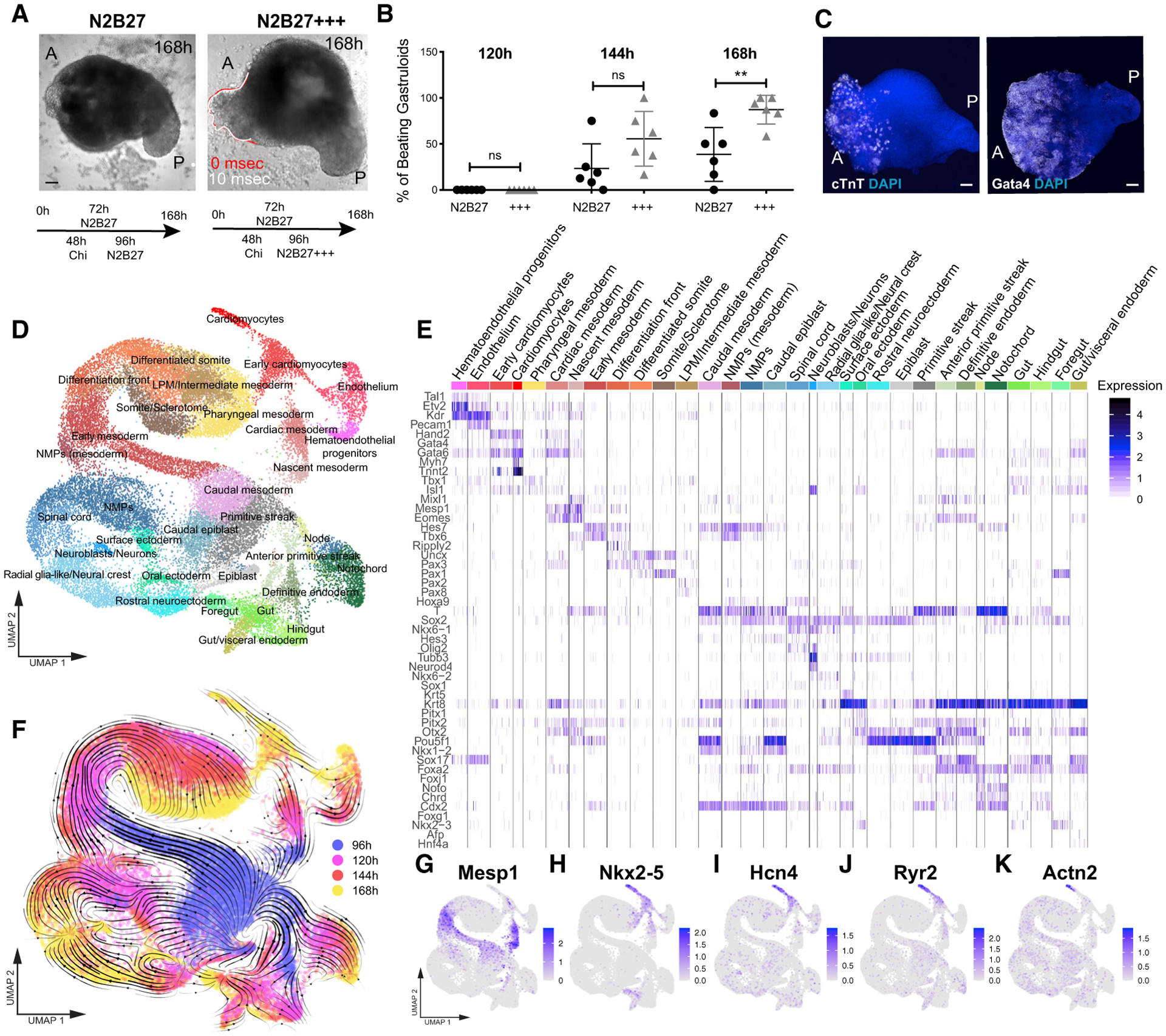
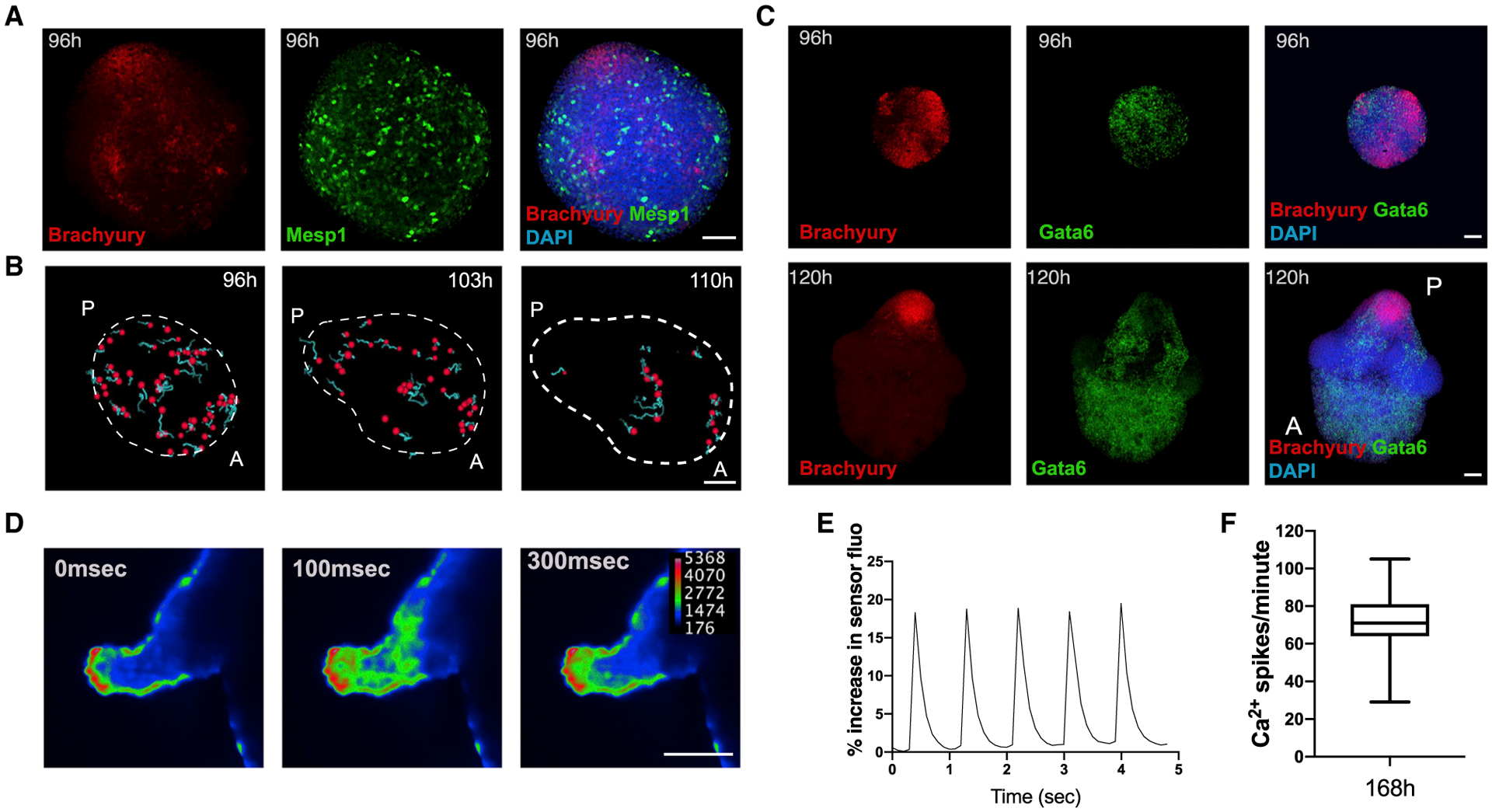
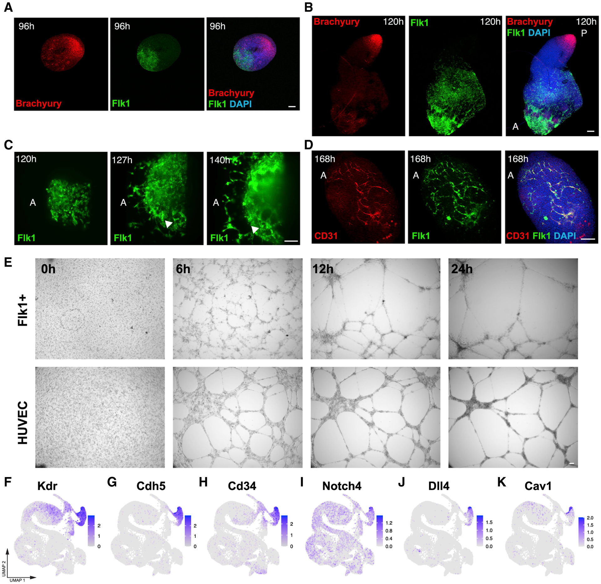
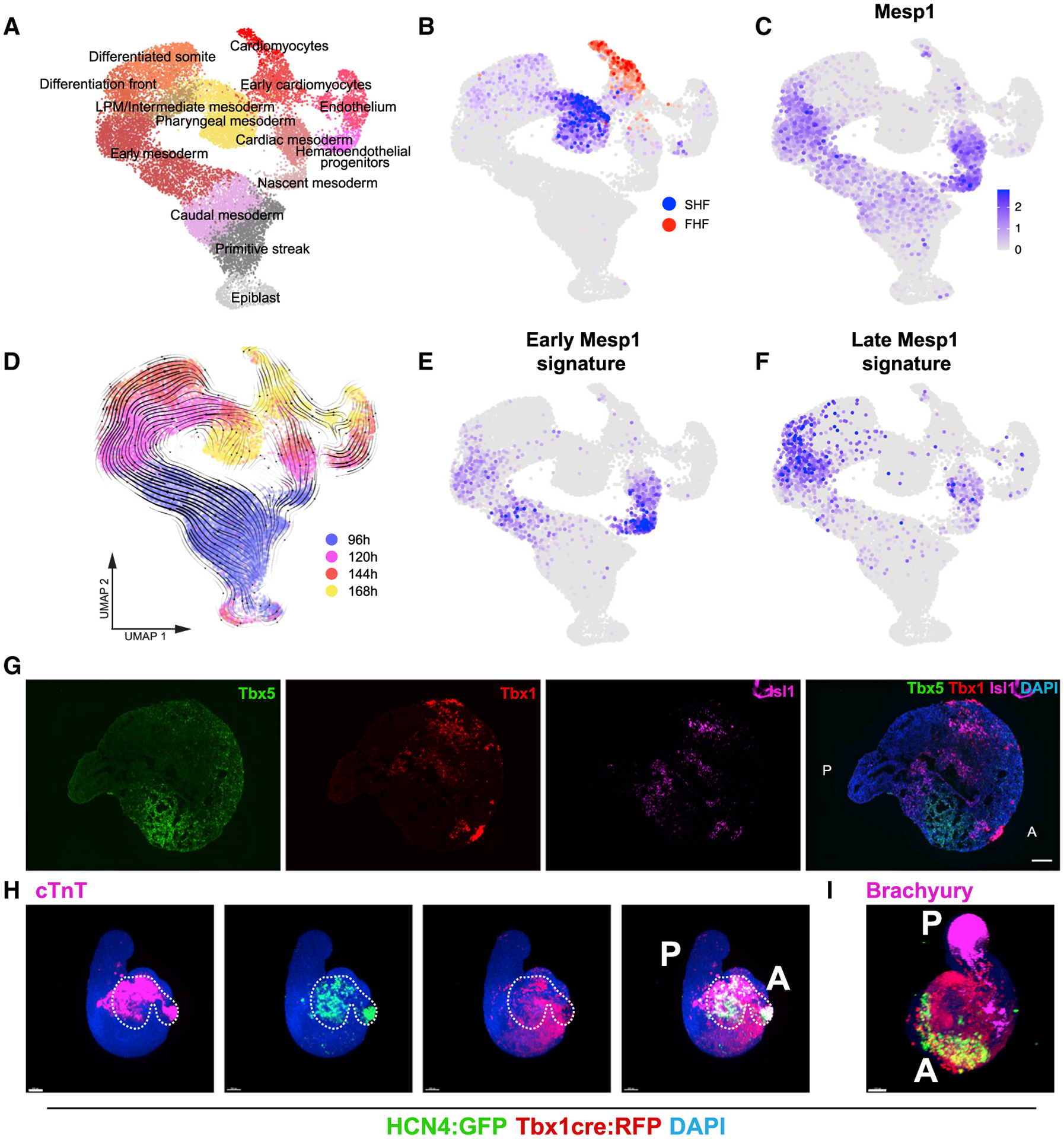
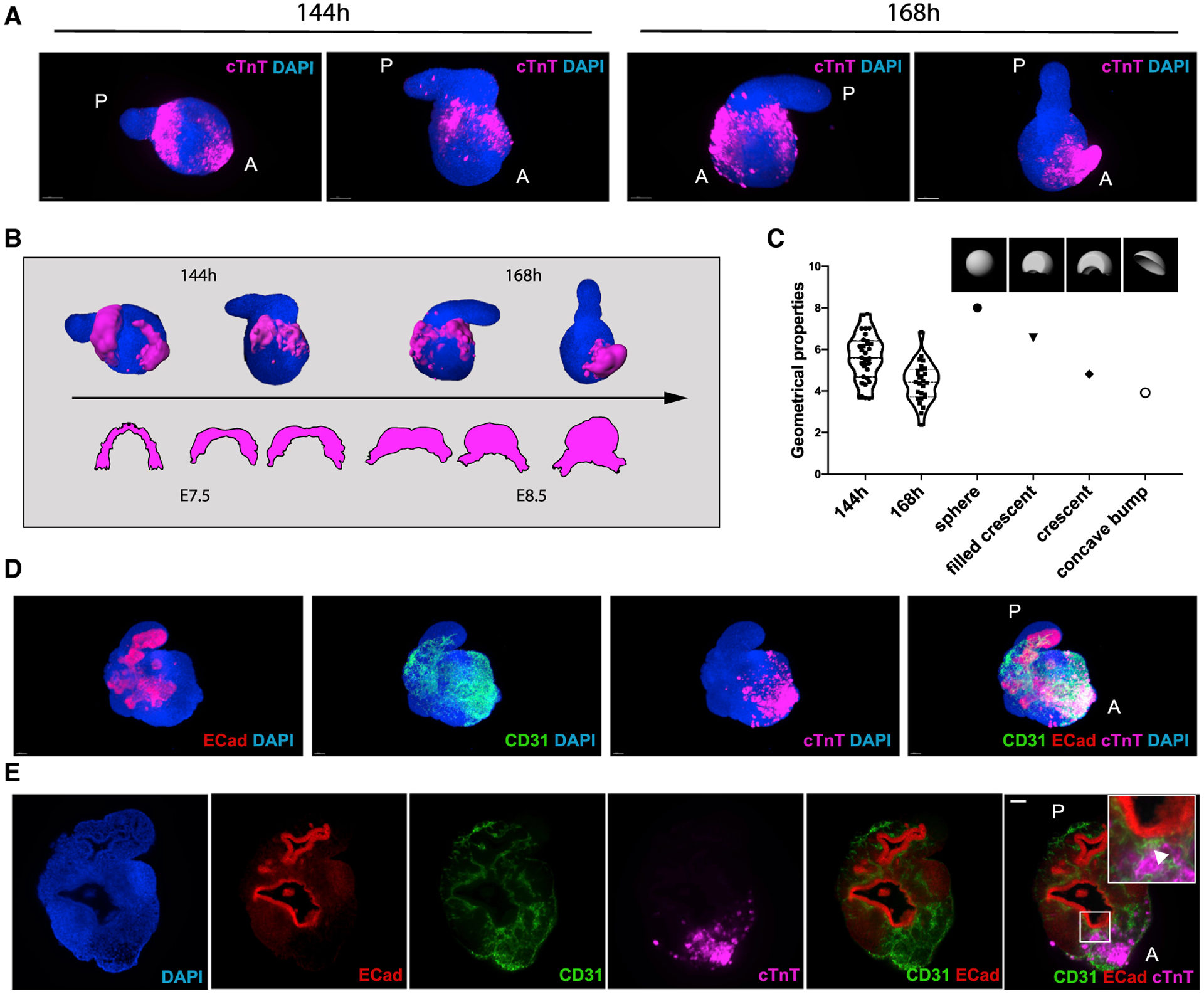
Organoids are powerful models for studying tissue development, physiology, and disease. However, current culture systems disrupt the inductive tissue-tissue interactions needed for the complex morphogenetic processes of native organogenesis. Here, we show that mouse embryonic stem cells (mESCs) can be coaxed to robustly undergo fundamental steps of early heart organogenesis with an in-vivo-like spatiotemporal fidelity. These axially patterned embryonic organoids (gastruloids) mimic embryonic development and support the generation of cardiovascular progenitors, including first and second heart fields. The cardiac progenitors self-organize into an anterior domain reminiscent of a cardiac crescent before forming a beating cardiac tissue near a putative primitive gut-like tube, from which it is separated by an endocardial-like layer. These findings unveil the surprising morphogenetic potential of mESCs to execute key aspects of organogenesis through the coordinated development of multiple tissues. This platform could be an excellent tool for studying heart development in unprecedented detail and throughput.

Keywords: 3D cardiac tissue; cardiac organoid; cardiogenesis; development; embryonic organoids; heart; in vitro organogenesis.


Conflict of interest statement

Declaration of Interests A.B. is part of Viventis Microscopy Sàrl that has commercialized the LS1 light-sheet microscope used in this study for time-lapse imaging of gastruloids. The EPFL (with Cambridge Enterprise Limited) has filed for patent protection (PCT/GB2019/052668) on the embryoid technology described herein, and M.P.L. and G.R. are named as inventors on the patent.

Figures

Figure 1.
Figure 1.. Embryonic Organoids Recapitulate Heart Organogenesis
(A) Gastruloids form a beating portion on their anterior side at 168 h. White and red lines highlight the displacement of the beating domain over 10 ms. (B) Frequency of beating structures at different time points in n = 6 independent experiments. (C) Immunofluorescence for Gata4 and cTnT on gastruloids at 168 h. (D) UMAP plot showing clusters obtained from 30,496 cells isolated from gastruloids at 96 h, 120 h, 144 h, and 168 h. n = 2 replicates per time point. (E) Heatmap showing the expression of canonical cell type markers illustrating the cell type diversity in (D). (F) Overlay of the time points analyzed (hours after aggregation) with the trajectories inferred from RNA velocity, projected on the uniform manifold approximation and projection (UMAP) in (D). (G–K) Expression of typical cardiac genes. Scale bars, 100 μm. A, anterior; P, posterior. See also Figures S1 and S2.
Figure 2.
Figure 2.. Early Cardiac Development in Embryonic Organoids Mimics Embryonic Development
(A) Spatial localization of Mesp1+ cells (stained for GFP) at 96 h as compared to Brachyury expression. (B) Tracking of Mesp1+ cells with live light-sheet imaging from 96 to 110 h. Red, tracked cells; light blue, cell tracks forward. (C) Spatial localization of Gata6+ cells at 96 and 120 h compared to Brachyury expression. (D) Calcium imaging of gastruloids at 168 h using a Cal-520 AM calcium dye and light-sheet microscopy. (E) Representative spiking profile. (F) Spiking frequency of the gastruloid cardiac portion at 168 h for n = 21 gastruloids. Data are represented as mean ± whiskers from min to max. Scale bars, 100 μm. See also Figure S2.
Figure 3.
Figure 3.. Development of a Vascular-like Network
(A and B) Spatial localization of Flk1+ cells at (A) 96 and (B) 120 h compared to Brachyury expression. Image in (B) is a composite of two separate images. (C) Light-sheet live imaging of the anterior portion of Flk1-GFP gastruloids from 120 to 144 h, highlighting the formation of a vascular-like network. (D) The Flk1+ vascular-like network is positive for CD31. (E) Angiogenesis assay showing tube formation in isolated Flk1+ cells compared to HUVECs. (F–K) UMAP plots showing the expression of endothelial marker genes (F–H) and genes related to endothelial cell maturation and morphogenesis (I–K) in the scRNA-seq dataset. Scale bars, 100 μm. See also Figure S3.
Figure 4.
Figure 4.. Embryonic Organoids Form a First and Second Heart Field
(A) UMAP plot showing clusters lying on the trajectories from epiblast to heart fields. n = 2 replicates per time point. (B) Localization of FHF on the UMAP in (A) based on expression of Tbx5, Hcn4, Nkx2–5, Hand1, and SHF based on the expression of Tbx1, Six1, and Six2. (C) Expression of Mesp1. (D) Overlay of time points and RNA velocity projected on the UMAP in (A). (E and F) Gene signature scores for early (E) and late (F) Mesp1-expressing cells in vivo based on in vivo differentially expressed genes from Lescroart et al. (2014). The scores are only shown for Mesp1-positive cells. (G) RNAscope showing spatial localization of FHF and SHF domains in gastruloids at 168 h. Images are stitched tile scans. (H) Representative immunofluorescence pictures of Hcn4-GFP::Tbx1Cre-RFP gastruloids at 168 h, showing the presence of FHF and SHF progenitors in the anterior domain of gastruloids. The white dashed line highlights the position of the Hcn4-positive domain in all pictures. n = 48 gastruloids from 4 independent experiments. (I) Representative immunofluorescence picture of Hcn4-GFP::Tbx1Cre-RFP gastruloids at 168 h, showing Brachyury expression in the posterior region. n = 32 gastruloids from 2 independent experiments. Scale bars, 100 μm. See also Figures S3 and S4.
Figure 5.
Figure 5.. Embryonic Organoids Recapitulate Cardiac Morphogenesis
(A) Light-sheet imaging of cleared gastruloids from 144 to 168 h show an initial crescent-like domain that is condensed into a beating bud at 168 h. (B) Schematic illustrating the comparison between gastruloid stages of cardiac development and embryonic stages from cardiac crescent to linear heart tube. (C) Quantification of geometrical properties (spareness) of the gastruloid cardiac domains compared to those of defined artificial shapes, with n = 31 gastruloids at 144 h and n = 27 gastruloids at 168 h. (D and E) The cardiac domain is localized near the anterior epithelial gut-tube-like structure, separated by a CD31+ endocardial layer. Scale bars, 100 μm. See also Figure S4.

Comment in

References

    1. Andersen P, Tampakakis E, Jimenez DV, Kannan S, Miyamoto M, Shin HK, Saberi A, Murphy S, Sulistio E, Chelko SP, and Kwon C (2018). Precardiac organoids form two heart fields via Bmp/Wnt signaling. Nat. Commun 9, 3140. - PMC - PubMed
    1. Baillie-Johnson P, van den Brink SC, Balayo T, Turner DA, and Martinez Arias A (2015). Generation of aggregates of mouse embryonic stem cells that show symmetry breaking, polarization and emergent collective behaviour in vitro. J. Vis. Exp 53252. - PMC - PubMed
    1. Beccari L, Moris N, Girgin M, Turner DA, Baillie-Johnson P, Cossy A-C, Lutolf MP, Duboule D, and Arias AM (2018). Multi-axial self-organization properties of mouse embryonic stem cells into gastruloids. Nature 562, 272–276. - PubMed
    1. Bergen V, Lange M, Peidli S, Wolf FA, and Theis FJ (2020). Generalizing RNA velocity to transient cell states through dynamical modeling. Nat. Biotechnol Published online August 3, 2020. 10.1038/s41587-020-0591-3. - DOI - PubMed
    1. Bondue A, Tännler S, Chiapparo G, Chabab S, Ramialison M, Paulissen C, Beck B, Harvey R, and Blanpain C (2011). Defining the earliest step of cardiovascular progenitor specification during embryonic stem cell differentiation. J. Cell Biol 192, 751–765. - PMC - PubMed

Publication types