Review
. 2020 Nov;13(11):e007405.
doi: 10.1161/CIRCHEARTFAILURE.120.007405. Epub 2020 Nov 12.

Management of Acute Myocarditis and Chronic Inflammatory Cardiomyopathy: An Expert Consensus Document

Affiliations
Review

Management of Acute Myocarditis and Chronic Inflammatory Cardiomyopathy: An Expert Consensus Document

Enrico Ammirati et al. Circ Heart Fail. 2020 Nov.

Abstract

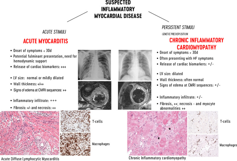
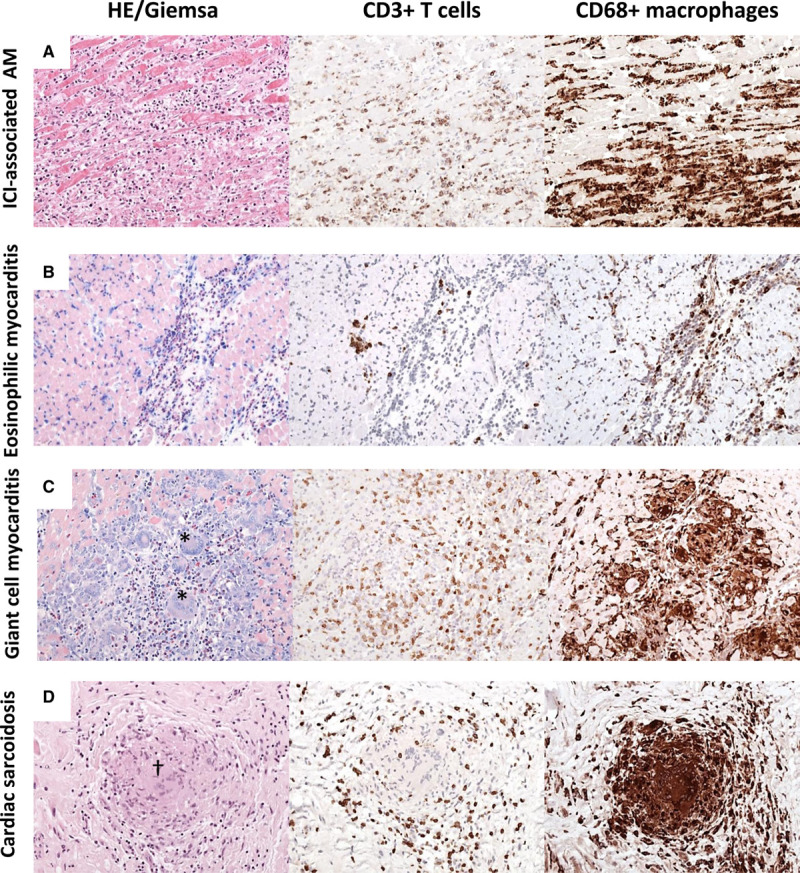
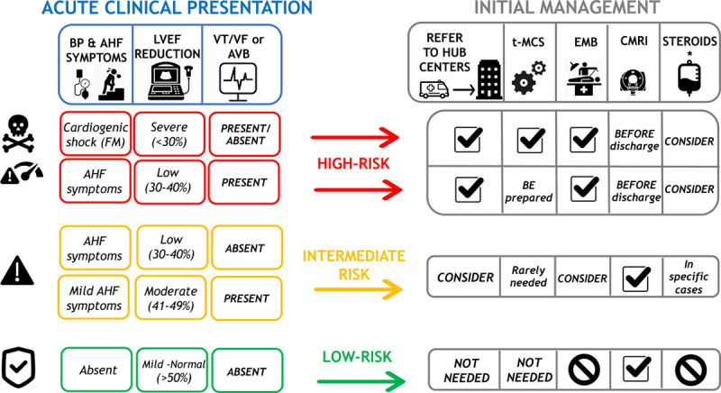
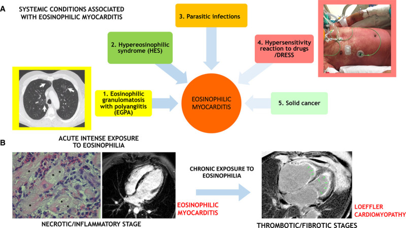
Myocarditis is an inflammatory disease of the heart that may occur because of infections, immune system activation, or exposure to drugs. The diagnosis of myocarditis has changed due to the introduction of cardiac magnetic resonance imaging. We present an expert consensus document aimed to summarize the common terminology related to myocarditis meanwhile highlighting some areas of controversies and uncertainties and the unmet clinical needs. In fact, controversies persist regarding mechanisms that determine the transition from the initial trigger to myocardial inflammation and from acute myocardial damage to chronic ventricular dysfunction. It is still uncertain which viruses (besides enteroviruses) cause direct tissue damage, act as triggers for immune-mediated damage, or both. Regarding terminology, myocarditis can be characterized according to etiology, phase, and severity of the disease, predominant symptoms, and pathological findings. Clinically, acute myocarditis (AM) implies a short time elapsed from the onset of symptoms and diagnosis (generally <1 month). In contrast, chronic inflammatory cardiomyopathy indicates myocardial inflammation with established dilated cardiomyopathy or hypokinetic nondilated phenotype, which in the advanced stages evolves into fibrosis without detectable inflammation. Suggested diagnostic and treatment recommendations for AM and chronic inflammatory cardiomyopathy are mainly based on expert opinion given the lack of well-designed contemporary clinical studies in the field. We will provide a shared and practical approach to patient diagnosis and management, underlying differences between the European and US scientific statements on this topic. We explain the role of histology that defines subtypes of myocarditis and its prognostic and therapeutic implications.

Keywords: cardiac magnetic resonance imaging; endomyocardial biopsy; inflammatory cardiomyopathy; myocarditis; viruses.


Conflict of interest statement

Dr Adler is a consultant for Abbott, Abiomed, AstraZeneca, Endotronix, Ionis, Medtronic, and Novartis, is on the board of directors of Genstem Therapeutics, and is a shareholder of Rocket Pharmaceuticals. Dr Brambatti is an employee at Ionis Pharmaceuticals. Dr Moslehi has served on advisory boards for Pfizer, Novartis, Bristol-Myers Squibb, Deciphera, Audentes Pharmaceuticals, Nektar, Takeda, Ipsen, Myokardia, AstraZeneca, GlaxoSmithKline, Intrexon, and Regeneron and has been supported by National Institutes of Health grants R56 HL141466 and R01 HL141466. Dr Tschöpe is a consultant at Cardiotropic Labs, Miami, FL. Dr Camici is a consultant at Servier. The other authors report no conflicts.

Figures

Figure 1.
Figure 1.
Characteristic features of lymphocytic acute myocarditis and chronic inflammatory cardiomyopathy. Left, Imaging features of acute myocarditis: chest radiograph of a patient admitted for chest pain and suspected acute myocarditis with no enlargement of the cardiac silhouette and cardiac magnetic resonance imaging (CMRI) showing normal left ventricular (LV) volume and significantly increased cardiac mass with diffuse high signal in T2-weighted images (arrows) suggesting diffuse edema. Histology shows acute lymphocytic myocarditis with myocyte necrosis and diffuse mononuclear cell infiltrates by hematoxylin-eosin and immunohistological stain on CD3+ T cells and CD68+ macrophages, compatible with an active myocarditis based on Dallas criteria (magnitude ×200). Right, Imaging features of chronic lymphocytic cardiomyopathy: chest radiograph of a patient admitted with heart failure (HF) symptoms, showing enlargement of cardiac silhouette; at CMRI, the LV is dilated, with normal thickness and focal areas of high signal intensity at T2-weighted images suggesting localized edema (arrows). At histology, chronic inflammatory cardiomyopathy typically presents fibrosis (*) within areas with inflammatory cellular infiltrates and myocyte abnormalities (magnitude ×200). CD indicates cluster of differentiation.
Figure 2.
Figure 2.
Different patterns of myocardial inflammation demonstrated by histological and immunohistological stainings on endomyocardial biopsy. A, Immune checkpoint inhibitor (ICI)–associated acute myocarditis (AM) frequently reveals diffuse mononuclear infiltrates composed of CD3+ T cells and CD68+ macrophages. Based on hematoxylin-eosin (HE) and immunohistological stainings, ICI-associated myocarditis resembles a diffuse lymphocytic myocarditis. B, In eosinophilic myocarditis, prominent inflammatory cells are eosinophilic granulocytes (Giemsa) and macrophages. C, Giant cell myocarditis is characterized by large mononucleated infiltrates with the presence of giant cells (*) and eosinophils (Giemsa). D, Cardiac sarcoidosis can be differentiated from giant cell myocarditis by the presence of granuloma (†) and absence of necrotic myocytes (magnitude, ×200 in all pictures). CD indicates cluster of differentiation.
Figure 3.
Figure 3.
Proposed risk-based approach to acute myocarditis. Left, Clinical features that characterize high (red boxes), intermediate (orange boxes), or low (green boxes) risk are summarized, according to the presence of low blood pressure (BP) and severity of acute heart failure (AHF), initial left ventricular ejection fraction (LVEF) on first echocardiogram, and ECG (presence of ventricular tachycardia [VT] or ventricular fibrillation [VF] or advanced atrioventricular block [AVB]). Right, How these risk features may influence patient management in terms of referral to expert centers, temporary mechanical circulatory support (t-MCS), need for endomyocardial biopsy (EMB) or cardiac magnetic resonance imaging (CMRI), and consideration for steroid treatment. Tag sign indicates recommended actions. No symbol indicates not recommended. *Immunosuppression with intravenous steroids may be considered and often used in patients with fulminant myocarditis; however, clinical studies that demonstrate their efficacy are lacking.
Figure 4.
Figure 4.
Eosinophilic myocardial injury: associated conditions and transition from acute myocarditis to inflammatory cardiomyopathy. A, Eosinophilic myocarditis can be idiopathic or associated with a systemic disorder. The associated conditions can be (1) eosinophilic granulomatosis with polyangiitis (EGPA), which is often associated with asthma, pulmonary nonfixed infiltrates (arrows on a chest computed tomographic scan image in the yellow inset), and paranasal sinus abnormalities; (2) hypereosinophilic syndromes (HES) characterized by persistent peripheral eosinophilia (≥1.5×10⁹/L for over 6 mo), which can be a complex idiopathic form or a myeloproliferative variant like the clonal form associated with FIP1L1/PDGFRA fusion gene; (3) parasitic infections; (4) hypersensitivity reactions to drugs and drug reaction with eosinophilia and systemic symptoms (DRESS) that are generally characterized by fever and diffuse skin rush (like in the patient with DRESS showed in the rose inlet), with frequent delay onset after drug initiation (up to 2–6 wk); and rarely, (5) solid tumors. While in the acute phase, the eosinophilic myocarditis is the main determinant of prognosis, the associated conditions can be the major determinants of prognosis in the mid and long term. B, An acute intense exposure to eosinophilia can cause an acute eosinophilic myocarditis (left), which in some case can be described as necrotizing due to extensive areas of the cardiomyocyte necrosis (*) caused by diffuse eosinophilic infiltrates on endomyocardial biopsy histology. The acute inflammatory phase can cause subendocardial and transmural injuries, identified by late gadolinium enhancement on cardiac magnetic resonance imaging (CMRI). If the eosinophilic exposure persists, eosinophilic injury evolves to a thrombotic and fibrotic stage, with diffuse subendocardial fibrosis with apical thrombi (green arrows), as identified by CMRI; the latter are characteristic features of Loeffler cardiomyopathy (right).

Comment in

References

    1. Cooper LT., JrMyocarditis. N Engl J Med. 2009;360:1526–1538. doi: 10.1056/NEJMra0800028 - PMC - PubMed
    1. Caforio AL, Pankuweit S, Arbustini E, Basso C, Gimeno-Blanes J, Felix SB, Fu M, Heliö T, Heymans S, Jahns R, et al. ; European Society of Cardiology Working Group on Myocardial and Pericardial Diseases. Current state of knowledge on aetiology, diagnosis, management, and therapy of myocarditis: a position statement of the European Society of Cardiology working group on myocardial and pericardial diseases. Eur Heart J. 2013;34:2636–48, 2648a. doi: 10.1093/eurheartj/eht210 - PubMed
    1. Richardson P, McKenna W, Bristow M, Maisch B, Mautner B, O’Connell J, Olsen E, Thiene G, Goodwin J, Gyarfas I, et al. Report of the 1995 World Health Organization/International Society and Federation of Cardiology task force on the definition and classification of cardiomyopathies. Circulation. 1996;93:841–842. doi: 10.1161/01.cir.93.5.841 - PubMed
    1. Corrado D, Basso C, Thiene G. Sudden cardiac death in young people with apparently normal heart. Cardiovasc Res. 2001;50:399–408. doi: 10.1016/s0008-6363(01)00254-1 - PubMed
    1. Harmon KG, Drezner JA, Maleszewski JJ, Lopez-Anderson M, Owens D, Prutkin JM, Asif IM, Klossner D, Ackerman MJ. Pathogeneses of sudden cardiac death in national collegiate athletic association athletes. Circ Arrhythm Electrophysiol. 2014;7:198–204. doi: 10.1161/CIRCEP.113.001376 - PubMed