

. 2020 Nov 12;13(11):CIRCHEARTFAILURE120007636.
doi: 10.1161/CIRCHEARTFAILURE.120.007636.
Online ahead of print.
Virus-Negative Myopericarditis in Human Coronavirus Infection: Report From an Autopsy Series
Affiliations
- PMID: 33176456
- PMCID: PMC7673636
- DOI: 10.1161/CIRCHEARTFAILURE.120.007636
Item in Clipboard
Virus-Negative Myopericarditis in Human Coronavirus Infection: Report From an Autopsy Series
Circ Heart Fail.
.
Abstract
Supplemental Digital Content is available in the text.
Keywords: autopsy; coronavirus infections; heart failure; hypertension; stroke volume.
Conflict of interest statement
None.
Figures
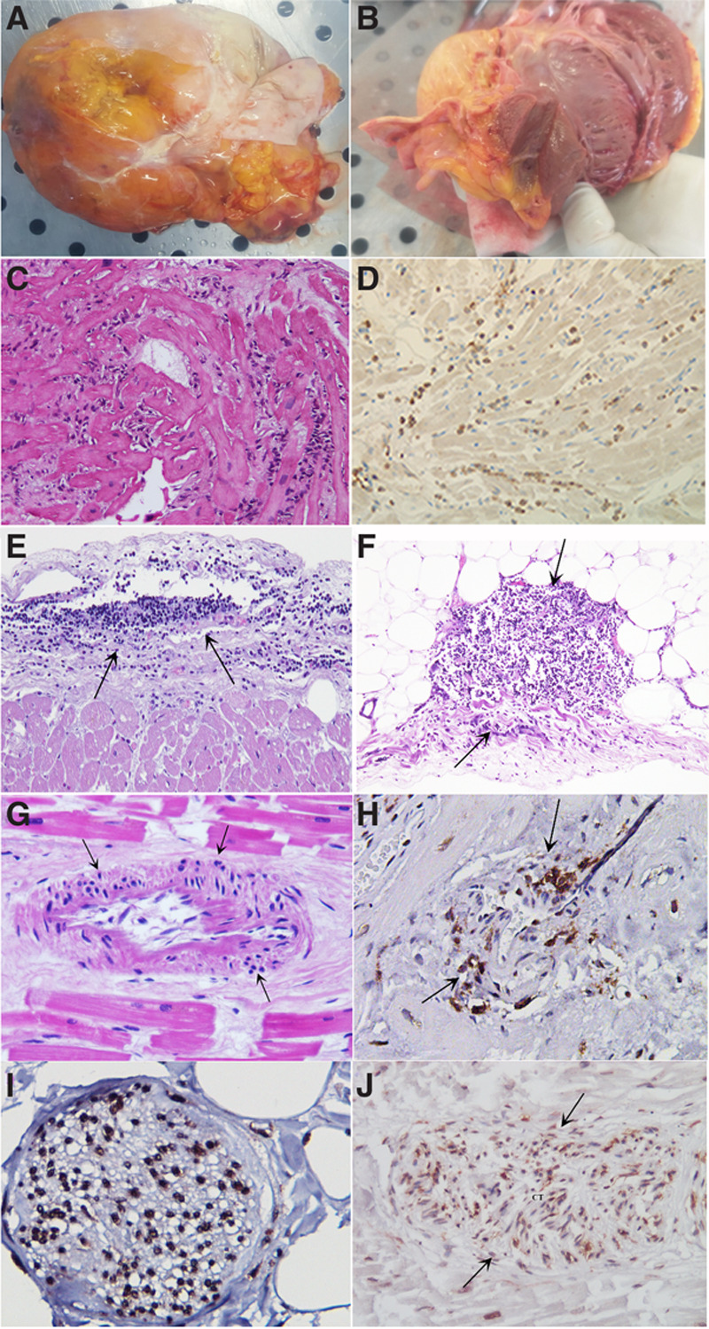
Cardiac postmortem findings in 9 patients affected by coronavirus disease 2019 (COVID-19) infection.
A, Overweight heart (450 g) in a patient with coronavirus infection. Plaque-like fibrous whitish thickened pericardium denoting pericarditis. B, Grossly, the heart was dilated, and the ventricular myocardium was flabby and pale with thickened walls. C, Lymphocytic myocarditis (hematoxlin and eosin, 200×). D, Immunohistochemistry for cluster differentiation68 showing positive macrophages in the inflamed myocardium. E and F, Lymphocytic pericarditis (hematoxlin and eosin, 200×). G, Necrotizing vasculitis of an intramural coronary artery (hematoxlin and eosin, 200×). H, Immunohistochemistry for Cluster differentiation45Ro suggesting lymphocytic infiltration and necrosis of an intramural coronary artery (200×). I, Lymphocytic infiltration of a subepicardial ganglion (cluster differentiation45Ro, 400×). J, Severe infiltration with damage of a section of conduction tissue (hematoxlin and eosin, 200×).
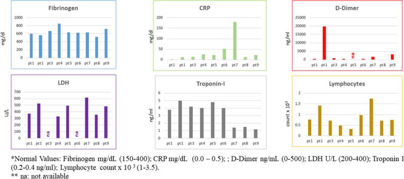
Biomarker data of 9 patients (pts) with coronavirus infection. The levels of fibrinogen, CRP, d -dimer, lactate dehydrogenase (LDH), troponin I, and lymphocytes were evaluated by diagnostic tools in 9 pts with coronavirus infection. CRP indicates C-reactive protein; and na, not available.
References
-
- Sala S, Peretto G, Gramegna M, Palmisano A, Villatore A, Vignale D, De Cobelli F, Tresoldi M, Cappelletti AM, Basso C, et al. Acute myocarditis presenting as a reverse Tako-Tsubo syndrome in a patient with SARS-CoV-2 respiratory infection. Eur Heart J. 2020;41:1861–1862. doi: 10.1093/eurheartj/ehaa286 - PMC - PubMed
LinkOut - more resources
Full Text Sources
