. 2020 Nov 11;51(1):136.
doi: 10.1186/s13567-020-00865-y.

Tomatidine inhibits porcine epidemic diarrhea virus replication by targeting 3CL protease

Affiliations

Tomatidine inhibits porcine epidemic diarrhea virus replication by targeting 3CL protease

Pengcheng Wang et al. Vet Res. .

Abstract

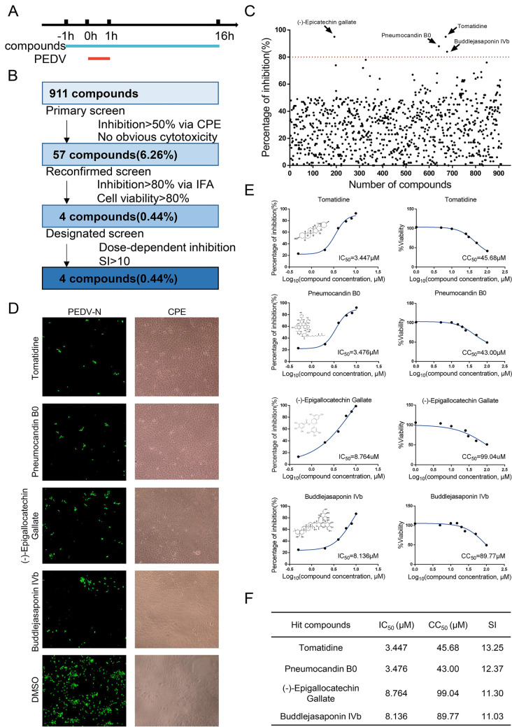
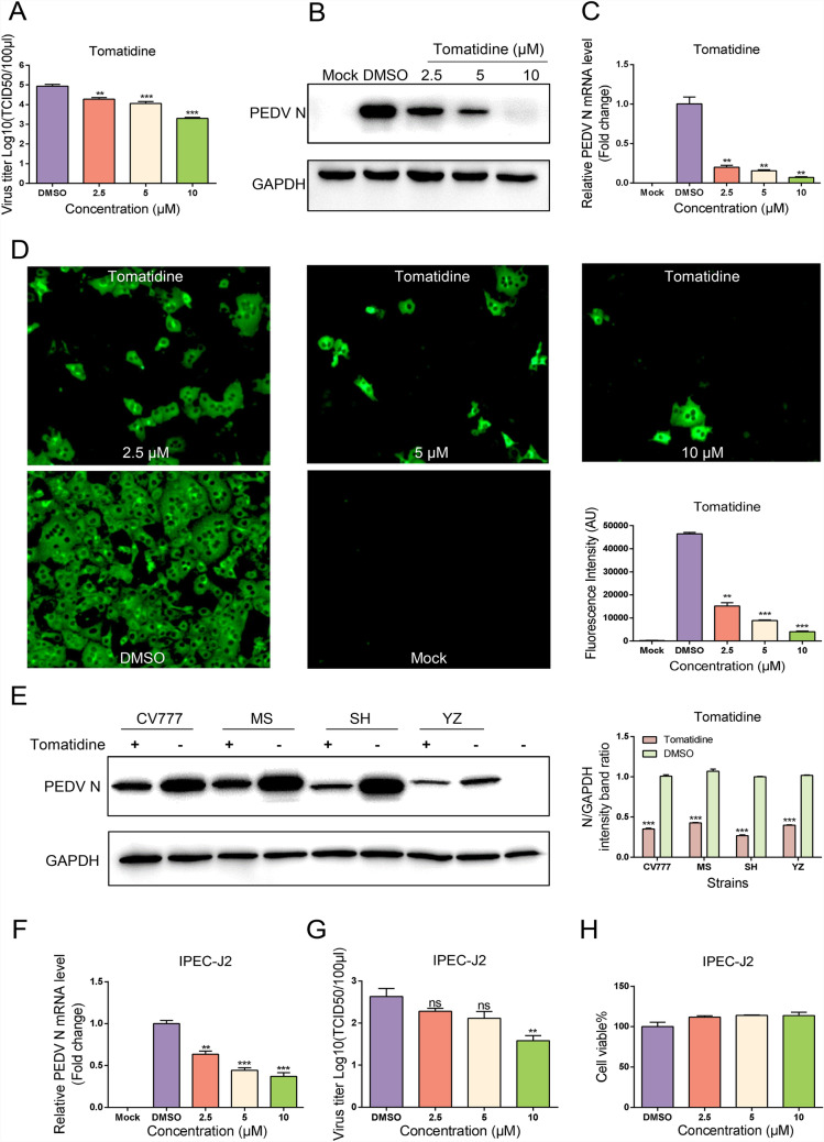
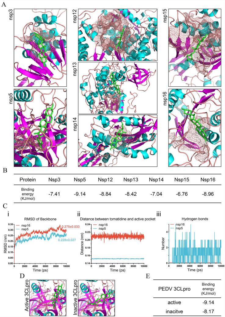
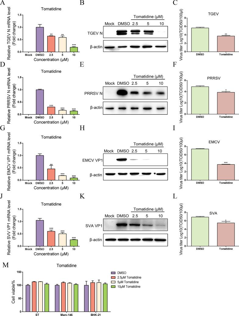
Porcine epidemic diarrhea virus (PEDV) causes lethal diarrhea in suckling piglets, leading to severe economic losses worldwide. There is an urgent need to find new therapeutic methods to prevent and control PEDV. Not only is there a shortage of commercial anti-PEDV drugs, but available commercial vaccines fail to protect against highly virulent PEDV variants. We screened an FDA-approved library of 911 natural products and found that tomatidine, a steroidal alkaloid extracted from the skin and leaves of tomatoes, demonstrates significant inhibition of PEDV replication in Vero and IPEC-J2 cells in vitro. Molecular docking and molecular dynamics analysis predicted interactions between tomatidine and the active pocket of PEDV 3CL protease, which were confirmed by fluorescence spectroscopy and isothermal titration calorimetry (ITC). The inhibiting effect of tomatidine on 3CL protease was determined using cleavage visualization and FRET assay. Tomatidine-mediated blocking of 3CL protease activity in PEDV-infected cells was examined by western blot detection of the viral polyprotein in PEDV-infected cells. It indicates that tomatidine inhibits PEDV replication mainly by targeting 3CL protease. In addition, tomatidine also has antiviral activity against transmissible gastroenteritis virus (TGEV), porcine reproductive and respiratory syndrome virus (PRRSV), encephalo myocarditis virus (EMCV) and seneca virus A (SVA) in vitro. These results may be helpful in developing a new prophylactic and therapeutic strategy against PEDV and other swine disease infections.

Keywords: 3CL protease; PEDV replication; tomatidine.


Conflict of interest statement

None of the authors have any possible conflicts of interest.

Figures

Figure 1
Figure 1
Screening protocol for PEDV inhibitors. A Screening procedure. Vero cells were treated with 10 μM compound for 1 h, then infected with PEDV (0.01 MOI) for 1 h. The cells were washed with PBS, then incubated in medium containing 10 μM compound for another 15 h. B Screening process flowchart. The criteria for passing the primary screening were that the compound must have no apparent cytotoxicity and must reduce CPE by at least 50% compared with the DMSO treatment. The criteria for passing the secondary screening were that the compound must leave cells at least 80% viable and inhibit PEDV by more than 80%. Compounds that passed the third screen inhibited PEDV in a dose-dependent manner and had a selective index (SI) higher than 10. C Each dot represents the percent inhibition of each compound. The dots located above the dotted line indicate 80% or greater inhibition. D IFA of infected cells treated with one of the four designated compounds. PEDV N-protein is colored green; a bright field shows CPE. E IC50 and CC50 curves of the four designated compounds. Cell viability is calculated as a percentage of the viability in the cells treated with the compounds divided by that in the DMSO-treated cells. The structure of each compound is inset. F SIs of the four designated compounds.
Figure 2
Figure 2
Identification of anti-PEDV activity of tomatidine in Vero and IPEC-J2 cells. Vero cells were pretreated with the indicated concentrations of tomatidine (AD) for 1 h, and then infected with PEDV for 1 h at 37 °C. The cells were washed with PBS, then incubated in fresh medium containing tomatidine for 16 h. DMSO served as the treatment control. A Culture supernatants were collected at the indicated time points for viral titration. Results are expressed as TCID50. Titers from three independent experiments are shown as mean ± SD (error bars). B Western blot of N-protein in cells infected with PEDV and treated with the indicated concentrations of tomatidine or DMSO, at 16 hpi. C Relative PEDV N mRNA levels, determined by qRT-PCR, and expressed relative to that in DMSO-treated cells. The internal loading control was GAPDH. D Light microscope and IFA images of Vero cells infected with PEDV and treated with tomatidine, at 16 hpi. Viral N-protein is green. E Western blot of N-protein in cells infected with different PEDV genotypes and treated with indicated concentrations of tomatidine or DMSO, at 16 hpi. F, G IPEC-J2 cells were pretreated with tomatidine for 1 h, and infected with PEDV for 1 h at 37 °C. After incubation for a total of 24 h in fresh medium containing tomatidine, the culture supernatants were collected at the indicated time points for viral TCID50 titration, and the relative PEDV N mRNA levels in the cells were determined by qRT-PCR as above. H Viability of cells pretreated with the indicated concentrations of tomatidine and incubated for 24 h in medium containing tomatidine. The results are from one of three independent experiments. Error bars represent the SD. The asterisks in the figures indicate significant differences (*P < 0.05; **P < 0.01; ***P < 0.001; ns: not significant).
Figure 3
Figure 3
Effect of tomatidine on the inactivation, attachment, entry, replication, and release of PEDV. A Inactivated assay. Four groups: A. PEDV (10 μM) + tomatidine, B. PEDV (0.01 MOI) + DMSO, C. tomatidine (10 μM), and D. DMSO, were prepared and incubated at 37 °C for 3 h. Group A was then mixed with group D, and group B was mixed with group C, and the mixtures were added into Vero cells seeded in 24-well plates. After incubation at 37 °C for another 1 h, culture supernatants were replaced with fresh DMEM and incubated for an additional 12 h. The cells were washed with PBS, and the mRNA levels of PEDV N and GAPDH in the cells were measured using qRT-PCR. B Virus attachment assay. C Virus internalization assay. D Virus replication assay. E Virus release assay. The results are from one of three independent experiments. Error bars represent the SD. The asterisks in the figures indicate significant differences (*P < 0.05; **P < 0.01; ***P < 0.001; ns: not significant).
Figure 4
Figure 4
In silico tomatidine targeted the active pocket of PEDV 3CL protease. A Docked conformations of tomatidine with PEDV nsp3 PLP2, nsp5 3CLpro, nsp12 RdRp, nsp13 NTP, nsp14 ExoN, nsp15 Nendo U, and nsp16 2′-o-methyltransferase. The compounds and proteins are represented as sticks and cartoons, respectively. The compounds are colored green. The proteins are colored according to their secondary structures (helix = blue, sheet = purple, loop = pink). The active sites of enzyme pockets are shown as a mesh. B The binding energy of the tomatidine–protein complex, calculated using Autodock, is listed. C Overall dynamic behaviors in the MD simulations. (i) RMSD of backbones of nsp5 (red) and nsp16 (blue); (ii) Distance between tomatidine and active pocket of nsp5 (red) and nsp16 (blue); (iii) Number of hydrogen bond interactions of tomatidine with nsp5 (red) and nsp16 (blue). D Docked conformations of tomatidine with PEDV 3CLpro or inactive 3CLpro. The compounds and proteins are represented as sticks and cartoons, respectively. The compounds are colored green. The proteins are colored according to their secondary structures (helix = blue, sheet = purple, loop = pink). The active sites of enzyme pockets are shown as a mesh. E The binding energy of the tomatidine–protein complex, calculated using Autodock, is listed.
Figure 5
Figure 5
Binding of tomatidine with 3CLpro detected by fluorescence quenching assay and ITC. A Expression and purification of recombinant 3CLpro. (i) BL-21 cells transformed with recombinant plasmids pET-32a-3CLpro were subjected to SDS-PAGE. Lane 1, BL21-pET-32a-3CLpro without IPTG induction; Lane 2, BL21-pET-32a with IPTG induction; Lane 3, BL21-pET-32a-3CLpro with IPTG induction; Lane 4, BL21-pET-32a-3CLpro with IPTG induction supernatant after ultrasonication; Lane 5, BL21-pET-32a-3CLpro with IPTG induction precipitation after ultrasonication. (ii) Recombination protein supernatant before (Lane 1) and after (Lane 2) purifications were subjected to SDS-PAGE. (iii) Purified protein was analyzed by Western blot. B Upper: Fluorescence emission spectra of different concentrations of tomatidine in the 3CLpro solutions at 50 mg/L; Lower: Stern–Volmer plot describes the 3CLpro quenching caused by association with quencher. C Representative thermodynamic profiles of tomatidine binding with the 3CLpro in solution, results of ITC measurements.
Figure 6
Figure 6
Tomatidine directly inhibited the activity of PEDV 3CL protease. A Vero cells cultured in 24-well plates were co-transfected with 250 ng of GFPnsp5/6 encoding plasmid, 250 ng of 3CLpro or inactive mutants expressing plasmid or empty vector. At 24 hpi, the cells were harvested and the cleaved fragment of GFPnsp5/6 was visualized using western blot. B Vero cells cultured in 24-well plates were co-transfected with 250 ng of GFPnsp5/6 encoding plasmid, 250 ng of 3CLpro expressing plasmid or empty vector. At 12 hpi, the culture supernatants were replaced with fresh DMEM containing the indicated concentrations (10, 20, and 30 μM) of tomatidine or DMSO. At 24 hpi, the cells were harvested and the cleaved fragment of GFPnsp5/6 was visualized using western blot. C PEDV 3CLpro was used at final concentrations of 0.5, 1, and 1.5 μM; and 10 μM (3CLpro substrate) was added to the protein in a black 96-well plate. PRRSV GP5 protein was used as a negative control. The mixtures were then further incubated at 37 °C for 20 min, and fluorescence was monitored at 340 nm excitation and 485 nm emissions every minute. The RFU were calculated by subtracting the mock from the fluorescence readings to eliminate the effect of background signals. D Tomatidine (25, 50 μM) or obacunone (50 μM, negative control) or DMSO was pre-incubated with 1 μM protease for 20 min at 37 °C, and 10 μM (3CLpro substrate) was added to the mixture in a black 96-well plate. The mixtures were then further incubated at 37 °C for 20 min, and fluorescence intensity was monitored at 340 nm excitation and 485 nm emissions every minute. RFU were calculated as above. E Vero cells were inoculated with PEDV (0.01 and 0.02 MOI) and 6 μM tomatidine. Cells were transfected with 250 ng pCAGGS-3CLpro as 3CLpro positive control. After incubation for 16 h, the cellular proteins were collected and the virus polyprotein was detected with western blot using anti-3CLpro mouse antibody, as previously described (20). Meanwhile, PEDV N-protein, GAPDH, and 3CLpro-Flag were detected with western blot using molecular antibodies against PEDV N-protein, GAPDH, and Flag. The ratio of nsp5-6 complex/N protein levels was depicted by integrated density analysis. The arrow indicates the location of nsp5-6 complex. All results are mean ± SD from three independent experiments performed in triplicate.
Figure 7
Figure 7
Tomatidine shows broad-spectrum antiviral activity against other swine disease viruses. TCID50, western blot, and qPCR were used to examine the inhibition activity of tomatidine against other swine disease viruses. Three designated concentrations of compounds were added to the culture medium (final concentrations were 2.5, 5, 10 μM, with no-observable cytotoxicity). DMSO was used as the negative control. TGEV (0.01 MOI), PRRSV (E), and EMCV (H)/SVA (K) were then used to infect ST, Marc-145, and BHK-21 cells, respectively, and samples were harvested at 24 h, 48 h, and 18 h. The TCID50 of TGEV (A), PRRSV (D), EMCV (G), and SVA (J) treated with 10 μM tomatidine or DMSO were calculated using the Reed-Muench method. The N-protein level of TGEV (B), PRRSV (E), EMCV (H), and SVA (K) were determined by western blot. Relative TGEV N (C), PRRSV N (F), EMCV VP1 (I), and SVA VP1 (L) mRNA levels, was determined by qRT-PCR, and expressed relative to that in DMSO-treated cells. M Viability of ST, Marc-145, and BHK-21 cells pretreated with the indicated concentrations of tomatidine and incubated for 24 h, 48 h, and 18 h, respectively in medium containing tomatidine. The results are from one of three independent experiments. The internal loading control was β-actin. Error bars represent the SD. The asterisks in the figures indicate significant differences (*P < 0.05; **P < 0.01; ***P < 0.001; ns = not significant).

References

    1. Jung K, Saif LJ. Porcine epidemic diarrhea virus infection: etiology, epidemiology, pathogenesis and immunoprophylaxis. Vet J. 2015;204(2):134–143. doi: 10.1016/j.tvjl.2015.02.017. - DOI - PMC - PubMed
    1. Lin CM, Saif LJ, Marthaler D, Wang Q. Evolution, antigenicity and pathogenicity of global porcine epidemic diarrhea virus strains. Virus Res. 2016;226:20–39. doi: 10.1016/j.virusres.2016.05.023. - DOI - PMC - PubMed
    1. Rota PA, Oberste MS, Monroe SS, Nix WA, Campagnoli R, Icenogle JP, Penaranda S, Bankamp B, Maher K, Chen MH, Tong S, Tamin A, Lowe L, Frace M, DeRisi JL, Chen Q, Wang D, Erdman DD, Peret TC, Burns C, Ksiazek TG, Rollin PE, Sanchez A, Liffick S, Holloway B, Limor J, McCaustland K, Olsen-Rasmussen M, Fouchier R, Gunther S, Osterhaus AD, Drosten C, Pallansch MA, Anderson LJ, Bellini WJ. Characterization of a novel coronavirus associated with severe acute respiratory syndrome. Science. 2003;300(5624):1394–1399. doi: 10.1126/science.1085952. - DOI - PubMed
    1. Assiri A, Al-Tawfiq JA, Al-Rabeeah AA, Al-Rabiah FA, Al-Hajjar S, Al-Barrak A, Flemban H, Al-Nassir WN, Balkhy HH, Al-Hakeem RF, Makhdoom HQ, Zumla AI, Memish ZA. Epidemiological, demographic, and clinical characteristics of 47 cases of Middle East respiratory syndrome coronavirus disease from Saudi Arabia: a descriptive study. Lancet Infect Dis. 2013;13(9):752–761. doi: 10.1016/s1473-3099(13)70204-4. - DOI - PMC - PubMed
    1. Yan R, Zhang Y, Li Y, Xia L, Guo Y, Zhou Q. Structural basis for the recognition of SARS-CoV-2 by full-length human ACE2. Science. 2020;367(6485):1444–1448. doi: 10.1126/science.abb2762. - DOI - PMC - PubMed

MeSH terms

LinkOut - more resources