. 2021 Jan 13;95(3):e01643-20.
doi: 10.1128/JVI.01643-20. Print 2021 Jan 13.

cDNA-Derived RNA Phage Assembly Reveals Critical Residues in the Maturation Protein of the Pseudomonas aeruginosa Leviphage PP7

Affiliations

cDNA-Derived RNA Phage Assembly Reveals Critical Residues in the Maturation Protein of the Pseudomonas aeruginosa Leviphage PP7

Eun Sook Kim et al. J Virol. .

Abstract

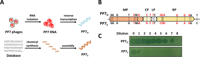
PP7 is a leviphage, with a single-stranded RNA genome, that infects Pseudomonas aeruginosa PAO1. A reverse genetic system for PP7 was previously created by using reverse-transcribed cDNA (PP7O) from a virion-derived RNA genome. Here, we have found that the PP7O cDNA contained 20 nucleotide differences from the PP7 genome sequence deposited in the database. We created another reverse genetic system exploiting chemically synthesized cDNA (PP7S) based on the database sequence. Unlike PP7O, which yielded infectious PP7 virions, PP7S-derived particles were incapable of plaque formation on PAO1 cells, which was restored in the PAO1 cells expressing the maturation protein (MP) from PP7O Using this reverse genetic system, we revealed two amino acid residues involved in the known roles of MP (i.e., adsorption and genome replication), fortuitously providing a lesson that the viral RNA genome sequencing needs functional verification, possibly by a reverse genetic system.IMPORTANCE The biological significance of RNA phages has been largely ignored, ironically, because few studies have focused on RNA phages. As an initial attempt to properly represent RNA phages in the phageome, we previously created, by using reverse-transcribed cDNA, a reverse genetic system for the small RNA phage PP7, which infects the opportunistic human pathogen Pseudomonas aeruginosa We report another system by using chemically synthesized cDNA based on the database genome that has 20 nucleotide differences from the previous cDNA. Investigation of those cDNA-derived phage virions revealed that two amino acids of the maturation protein are crucial for the normal phage lifecycle at different steps. Our study provides insight into the molecular basis for the RNA phage lifecycle and a lesson that the RNA genome sequencing needs to be carefully validated by cDNA-based phage assembly systems.

Keywords: PP7; Pseudomonas aeruginosa; RNA phage; cDNA; infectivity; maturation protein.


Figures

FIG 1
FIG 1
Construction of cDNA-derived RNA phage assembly systems. (A) Schematic representation of cDNA synthesis. PP7O was reverse transcribed from isolated PP7 genomic RNA (upper), and PP7S was chemically synthesized using the PP7 RNA genome sequence deposited in the public database (accession no. NC_001628) (bottom). (B) Sequence differences between PP7O and PP7S cDNA clones (Table 1). The different nucleotides between PP7O and PP7S are indicated by solid lines on the PP7 genome map, showing 4 coding regions: MP, maturation protein; CP, coat protein; LP, lysis protein; RP, RNA-dependent RNA polymerase (RdRp). Eleven nucleotide differences, without affecting the codons, are designated in red. (C) Phage production from PP7O and PP7S cDNA clones. The supernatant of the surrogate strain (PAK) harboring either PP7O or PP7S cDNA was diluted serially and spotted onto the susceptible strain (PAO1). The numbers indicate the dilution degree of the supernatant as −log10(fold dilution).
FIG 2
FIG 2
Phage production from the two cDNA clones. (A) Comparison of the RNA levels from the cDNA clones. Total cellular RNAs from PAK cells containing either PP7O (white) or PP7S (grey) cDNA were analyzed by RT-qPCR, with the amount of the rpoA mRNA as the control. (B) Transmission electron micrographs of PP7O- and PP7S-derived virions. Each virion sample was prepared for PP7O and PP7S, as described in Materials and Methods, and then subjected to negative staining with 2% uranyl acetate. Scale bars represent 50 or 100 nm. (C to E) Phage amounts were enumerated by counting the particle numbers, as shown in panel B (C), by measuring the RNA levels (D), and by investigating the coat protein (CP) levels (E). The CP (∼14-kDa) bands are designated by the arrowhead. C, 109 PFU of the PP7 phage samples. The numbers at the bottom represent the relative band intensity based on PP7O.
FIG 3
FIG 3
Identification of the residues critical for infectivity. (A) Schematic representation of the recombinant PP7 cDNA clones. A chimeric clone (WT) containing the ORF1 gene (MP) from PP7O (yellow) and the rest from PP7S (empty) was used as the template to create the 6 missense mutants (A125G, A150G, G380A, T872G, G1125A, and C1158G) and one silent mutant (G1162A) with the corresponding codon changes indicated. (B) Plaque formation of the mutant phages. The phage particles obtained from the cDNA clones in panel A were evaluated for their plaque formation ability on PAO1. Plaques were visualized after 16 h of incubation at 37°C.
FIG 4
FIG 4
Characterization of G1125(R349) and C1158(S360) mutations. (A) Potential RNA secondary structure of the regions around G1125 and C1158. RNA secondary structure of the PP7 genomic RNA was predicted using the RNA secondary structure prediction site (http://rna.tbi.univie.ac.at/forna/), and the corresponding regions are depicted with both G1125 and C1158, indicated in red. The codons (CGA for R349 and UCU for S360) are in the grey box. (B) Plaque formation of the mutant phages. The remaining point mutant phages at G1125 and C1158 (3 for each) and two silent transversion mutant phages (R349R and S360S) were prepared, and their plaque formation was analyzed by spotting assay as described for Fig. 1C. The numbers indicate the dilution degree of the supernatant as −log10 (fold dilution).
FIG 5
FIG 5
Effects on adsorption efficiency and RNA genome synthesis by R349 and S360 mutations. (A) Adsorption efficiency of the mutant phages. The phage mutants for the MP (R349Q, S360C, and R349Q/S360C double) as well as the PP7O- and the PP7S-derived phages were incubated with the wild-type (WT) PAO1 and the pilA mutant. Unbound phages in the supernatant were enumerated by plaque assay to calculate the adsorption efficiency. The numbers in each photograph indicate the numbers of the plaques, and the numbers at the bottom designate the calculated adsorption efficiency. (B) The genomic RNA levels immediately after infection by the mutant phages. PAO1 cells were infected by one of the mutant phages as described for panel A: S360C (yellow), R349Q (blue), R349Q/S360C double (green), PP7O (purple), and PP7S (orange) phages. The RNA levels from the infected cells were analyzed by RT-qPCR and the ΔΔCT values with the rpoA mRNA levels as the internal controls. A base-10 logarithmic (log) scale is used for the y axis with standard errors from three biological replicates.
FIG 6
FIG 6
Complementation of the mutants by maturation protein (MP). (A) Plaque formation on PAO1 cells expressing MP. Phage particles from PAK cells harboring PP7O or PP7S and two mutant [G1125A(R349Q) and C1158G(S360C)] cDNA clones and the MP-expressing plasmid (pJN105-MP) were spotted on PAO1 harboring pJN105 (left) and pJN105-MP (right). The numbers indicate the dilution degree of the supernatant as −log10(fold dilution). The plaques subjected to sequence determination are in the dotted box. (B) Sequence verification of the mutant phages. Phage RNAs that had been prepared from the phage plaques in panel A were subjected to RT-PCR. The partial nucleotide sequences of the PCR products are depicted in the electropherogram, and the reversion mutations (i.e., G1167C) observed for PP7S and C1158G(S360C) without (left) and with (right) MP expression are in red.

References

    1. Nolden T, Pfaff F, Nemitz S, Freuling CM, Hoper D, Muller T, Finke S. 2016. Reverse genetics in high throughput: rapid generation of complete negative strand RNA virus cDNA clones and recombinant viruses thereof. Sci Rep 6:23887. doi:10.1038/srep23887. - DOI - PMC - PubMed
    1. Racaniello VR, Baltimore D. 1981. Cloned poliovirus complementary DNA is infectious in mammalian cells. Science 214:916–919. doi:10.1126/science.6272391. - DOI - PubMed
    1. Frietze KM, Peabody DS, Chackerian B. 2016. Engineering virus-like particles as vaccine platforms. Curr Opin Virol 18:44–49. doi:10.1016/j.coviro.2016.03.001. - DOI - PMC - PubMed
    1. Palladini A, Thrane S, Janitzek CM, Pihl J, Clemmensen SB, de Jongh WA, Clausen TM, Nicoletti G, Landuzzi L, Penichet ML, Balboni T, Ianzano ML, Giusti V, Theander TG, Nielsen MA, Salanti A, Lollini P-L, Nanni P, Sander AF. 2018. Virus-like particle display of HER2 induces potent anti-cancer responses. Oncoimmunology 7:e1408749. doi:10.1080/2162402X.2017.1408749. - DOI - PMC - PubMed
    1. Zdanowicz M, Chroboczek J. 2016. Virus-like particles as drug delivery vectors. Acta Biochim Pol 63:469–473. doi:10.18388/abp.2016_1275. - DOI - PubMed

Publication types

MeSH terms