

Compensatory Movement Patterns Are Based on Abnormal Activity of the Biceps Brachii and Posterior Deltoid Muscles in Patients with Symptomatic Rotator Cuff Tears
- PMID: 33177479
- PMCID: PMC7899608
- DOI: 10.1097/CORR.0000000000001555
Compensatory Movement Patterns Are Based on Abnormal Activity of the Biceps Brachii and Posterior Deltoid Muscles in Patients with Symptomatic Rotator Cuff Tears
Abstract
Background: Abnormal movement patterns due to compensatory mechanisms have been reported in patients with rotator cuff tears. The long head of the biceps tendon may especially be overactive and a source of pain and could induce abnormal muscle activation in these patients. It is still unknown why some patients with a rotator cuff tear develop complaints and others do not.
Questions/purposes: (1) Which shoulder muscles show a different activation pattern on electromyography (EMG) while performing the Functional Impairment Test-Hand and Neck/Shoulder/Arm (FIT-HaNSA) in patients with a symptomatic rotator cuff tear compared with age-matched controls with an intact rotator cuff? (2) Which shoulder muscles are coactivated on EMG while performing the FIT-HaNSA?
Methods: This comparative study included two groups of people aged 50 years and older: a group of patients with chronic symptomatic rotator cuff tears (confirmed by MRI or ultrasound with the exclusion of Patte stage 3 and massive rotator cuff tears) and a control group of volunteers without shoulder conditions. Starting January 2019, 12 patients with a chronic rotator cuff tear were consecutively recruited at the outpatient orthopaedic clinic. Eleven age-matched controls (randomly recruited by posters in the hospital) were included after assuring the absence of shoulder complaints and an intact rotator cuff on ultrasound imaging. The upper limb was examined using the FIT-HaNSA (score: 0 [worst] to 300 seconds [best]), shoulder-specific instruments, health-related quality of life, and EMG recordings of 10 shoulder girdle muscles while performing a tailored FIT-HaNSA.
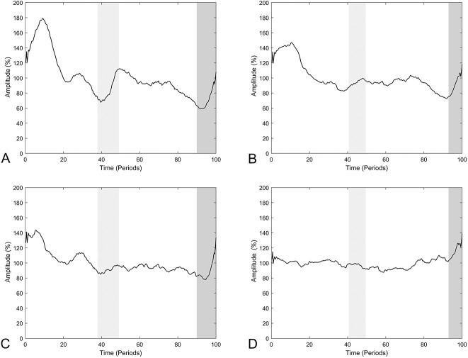
Results: EMG (normalized root mean square amplitudes) revealed hyperactivity of the posterior deltoid and biceps brachii muscles during the upward phase in patients with rotator cuff tears compared with controls (posterior deltoid: 111% ± 6% versus 102% ± 10%, mean difference -9 [95% confidence interval -17 to -1]; p = 0.03; biceps brachii: 118% ± 7% versus 111% ± 6%, mean difference -7 [95% CI -13 to 0]; p = 0.04), and there was decreased activity during the downward phase in patients with rotator cuff tears compared with controls (posterior deltoid: 89% ± 6% versus 98% ± 10%, mean difference 9 [95% CI 1 to 17]; p = 0.03; biceps brachii: 82% ± 7% versus 89% ± 6%, mean difference 7 [95% CI 0 to 14]; p = 0.03). The posterior deltoid functioned less in conjunction with the other deltoid muscles, and lower coactivation was seen in the remaining intact rotator cuff muscles in the rotator cuff tear group than in the control group.
Conclusion: Patients with a symptomatic rotator cuff tear show compensatory movement patterns based on abnormal activity of the biceps brachii and posterior deltoid muscles when compared with age-matched controls. The posterior deltoid functions less in conjunction with the other deltoid muscles, and lower coactivation was seen in the remaining intact rotator cuff muscles in the rotator cuff tear group than the control group.
Clinical relevance: This study supports the potential benefit of addressing the long head biceps tendon in the treatment of patients with a symptomatic rotator cuff tear. Moreover, clinicians might use these findings for conservative treatment; the posterior deltoid can be specifically trained to help compensate for the deficient rotator cuff.
Copyright © 2020 by the Association of Bone and Joint Surgeons.
Conflict of interest statement
All ICMJE Conflict of Interest Forms for authors and Clinical Orthopaedics and Related Research® editors and board members are on file with the publication and can be viewed on request. Each author certifies that neither he nor she, nor any member of his or her immediate family, has funding or commercial associations (consultancies, stock ownership, equity interest, patent/licensing arrangements, etc.) that might pose a conflict of interest in connection with the submitted article.
Figures
Comment in
-
CORR Insights®: Compensatory Movement Patterns Are Based on Abnormal Activity of the Biceps Brachii and Posterior Deltoid Muscles in Patients with Symptomatic Rotator Cuff Tears.Clin Orthop Relat Res. 2021 Feb 1;479(2):389-391. doi: 10.1097/CORR.0000000000001580. Clin Orthop Relat Res. 2021. PMID: 33475299 Free PMC article. No abstract available.
References
-
- Basmajian JV, De Luca CJ. Description and Analysis of the EMG Signal. In: Basmajian JV, ed. Muscles Alive: Their Functions Revealed by Electromyography. Williams & Wilkins; 1985.
-
- Boileau P, Ahrens PM, Hatzidakis AM. Entrapment of the long head of the biceps tendon: the hourglass biceps—a cause of pain and locking of the shoulder. J Shoulder Elbow Surg. 2004;13:249-257. - PubMed
-
- Burden AM, Trew M, Baltzopoulos V. Normalisation of gait EMGs: a re-examination. J Electromyogr Kinesiol. 2003;13:519-532. - PubMed
-
- Cioni R, Giannini F, Paradiso C, Battistini N, Navona C, Starita A. Sex differences in surface EMG interference pattern power spectrum. J Appl Physiol. 1994;77:2163-2168. - PubMed
Publication types
MeSH terms
LinkOut - more resources
Full Text Sources
Medical
