. 2020 Nov 11;10(1):19587.
doi: 10.1038/s41598-020-76350-5.

Dengue virus infection impedes megakaryopoiesis in MEG-01 cells where the virus envelope protein interacts with the transcription factor TAL-1

Affiliations

Dengue virus infection impedes megakaryopoiesis in MEG-01 cells where the virus envelope protein interacts with the transcription factor TAL-1

Atoshi Banerjee et al. Sci Rep. .

Abstract

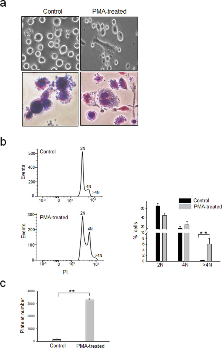
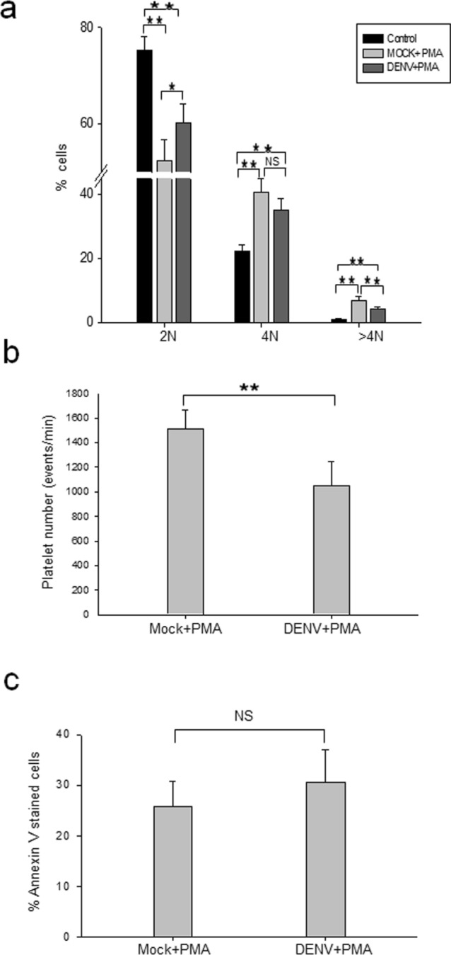
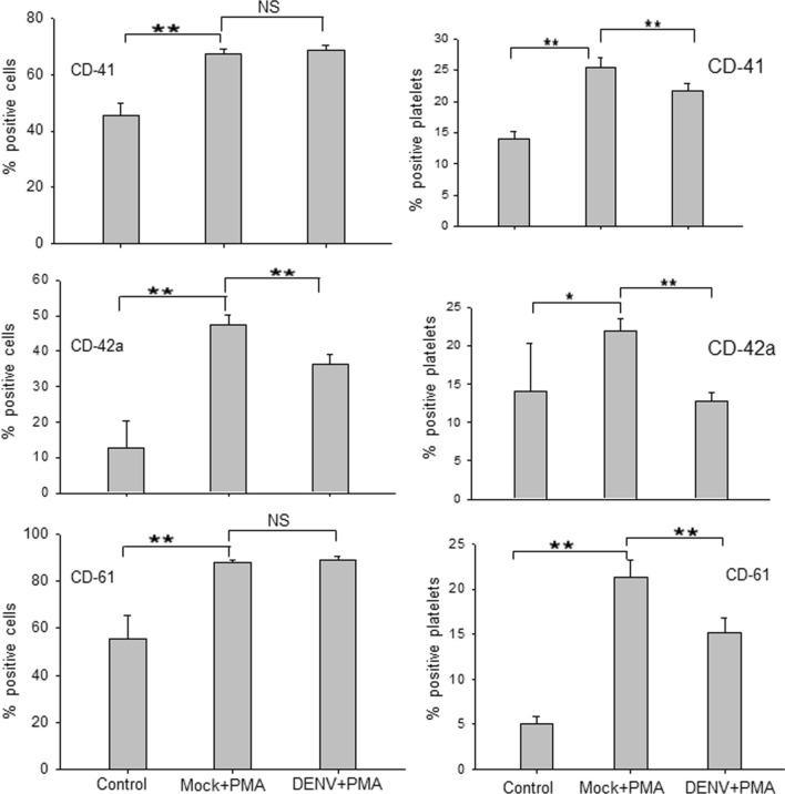
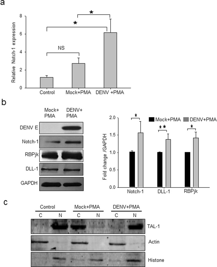
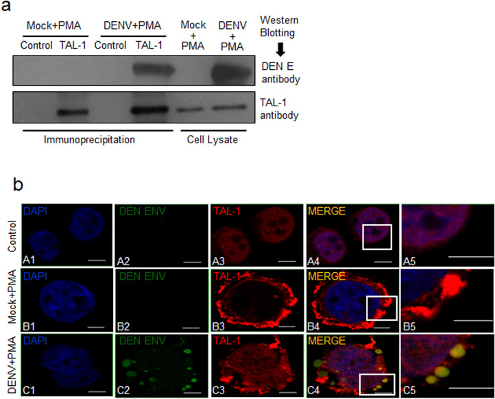
Dengue virus (DENV) infection causes dengue fever in humans, which can lead to thrombocytopenia showing a marked reduction in platelet counts, and dengue hemorrhagic fever. The virus may cause thrombocytopenia either by destroying the platelets or by interfering with their generation via the process of megakaryopoiesis. MEG-01 is the human megakaryoblastic leukemia cell line that can be differentiated in vitro by phorbol-12-myristate-13-acetate (PMA) treatment to produce platelet-like-particles (PLPs). We have studied DENV infection of MEG-01 cells to understand its effect on megakaryopoiesis and the generation of PLPs. We observed that DENV could infect only naive MEG-01 cells, and differentiated cells were refractory to virus infection/replication. However, DENV-infected MEG-01 cells, when induced for differentiation with PMA, supported an enhanced viral replication. Following the virus infection, the MEG-01 cells showed a marked reduction in the surface expression of platelet markers (CD41, CD42a, and CD61), a decreased polyploidy, and significantly reduced PLP counts. DENV infection caused an enhanced Notch signaling in MEG-01 cells where the virus envelope protein was shown to interact with TAL-1, a host protein important for megakaryopoiesis. These observations provide new insight into the role of DENV in modulating the megakaryopoiesis and platelet production process.


Conflict of interest statement

The authors declare no competing interests.

Figures

Figure 1
Figure 1
PMA treatment of MEG-01 cells induces differentiation and enhanced production of platelet-like particles. (a) MEG-01 cells were incubated for 3 days in RPMI 1640 medium (control) or in RPMI 1640 containing 50 ng/ml PMA (PMA-treated) and photographed at 20× resolution under an inverted light microscope (upper panels). Untreated control cells or PMA-treated MEG-01 cells were fixed with Methanol and subjected to May-Grünwald-Giemsa staining and visualized at 20× resolution under an inverted light microscope (lower panels). Experiment were repeated 3 times and representative images are shown. (b) MEG-01 cells treated as above were fixed and stained with PI and acquired by FACS for analyzing the cell cycle. The 2 N, 4 N, and > 4 N population were analyzed by FlowJo software. The left panel shows data from a typical experiment. The data in the right panel is from 3 replicates of the experiment. (c) MEG-01 cells were incubated in RPMI 1640 with or without PMA (50 ng/ml) for 5 days. The culture supernatant was collected and processed for PLP preparation. The PLPs were stained with CD41 antibody and analyzed by FACS. PLPs were identified by size and shape (side and forward scatter) where normal human platelets would fall and the presence of CD41 marker. Ten thousand events were recorded and platelet count was quantified by the rate (platelet events/min). The data is from 3 independent experiments.
Figure 2
Figure 2
DENV infection of MEG-01 cells. MEG-01 cells were mock-infected, or infected with DENV at a multiplicity of infection (MOI) 1, and the cells and culture supernatant were harvested on different days post-infection (pi). In parallel, cells were pre-treated with PMA 2 h before infection (PMA + DENV) or infected with DENV followed by PMA treatment (DENV + PMA). (a) The total RNA from cells was isolated on day 2 pi and relative levels of viral RNA determined by qRT-PCR using Gapdh transcripts for normalization. (b) The culture supernatants from virus-infected cells were collected on days 3 and 5 pi and DENV titers determined by FFU assays. All the experiments were done more than 3 times and data represent the mean ± standard deviation. Statistical analyses were done using the one-way analysis of variance (ANOVA). *p < 0.05 and **p < 0.005.
Figure 3
Figure 3
DENV infection of MEG-01 cells reduces polyploidy and production of PLPs. (a) MEG-01 cells were infected with DENV (MOI 1). DENV + PMA and Mock + PMA cells, along with naïve MEG-01 cells (untreated control) were incubated in culture medium at 37 °C for 5 days. Cells were fixed and stained with PI, and acquired by FACS for analyzing the cell cycle. The 2 N, 4 N, and > 4 N populations were analyzed by FlowJo software. (b) Culture supernatant from Mock + PMA and DENV + PMA treated cells were collected and processed for isolation of PLPs. The PLPs were stained with CD41 antibody and analyzed by FACS. PLPs were identified by size and shape (side and forward scatter) where the normal human platelets would fall and the presence of CD41 marker. Ten thousand events were recorded and platelet count was quantified by the rate (platelet events/min). (c) Cells harvested at day 5 pi were stained with Annexin V-alexa 488 antibody and acquired by FACS. Annexin V positive and negative cells were gated and analyzed. All the experiments were done more than 6 times and data represent the mean ± standard deviation. Statistical analyses were done using the one-way analysis of variance (ANOVA). *p < 0.05 and **p < 0.005.
Figure 4
Figure 4
DENV infection arrests surface marker expression acquired during MEG-01 cell differentiation. Naïve MEG-01 cells (control) were treated with PMA (Mock + PMA) or DENV-infected (MOI-1) cells were treated with PMA (DENV + PMA). Cells and PLPs were harvested at day 5 pi and stained with CD61-APC, CD42a-Alexa 488, and CD41-PE antibodies. Cells and platelets were determined based on SSC and FSC by FACS. For multicolor analysis, compensation was carried out using unstained and single-color samples and compensated voltages were applied to all the experimental samples. All the samples were acquired in the same compensated setting and analysis was done by FlowJo software. All the experiments were done more than 6 times and data represent the mean ± standard deviation. Statistical analyses were done using the one-way analysis of variance (ANOVA). *p < 0.05 and **p < 0.005, NS denotes difference not-significant.
Figure 5
Figure 5
Notch signaling in DENV-infected MEG-01 cells. Naïve MEG-01 cells (control) were treated with PMA (Mock + PMA), or DENV-infected (MOI-1) cells were treated with PMA (DENV + PMA). Cells were harvested at day 5 pi for isolating total RNA and preparation of cell lysate. (a) Expression of Notch-1 transcript relative to Gapdh expression is presented from 3 independent experiments. (b) The cell lysate was Western blotted for different proteins (left panel) and the band intensities were quantified by ImageJ software. Intensities from 3 or more blots were determined compared to GAPDH, and average intensities in DENV + PMA compared to Mock + PMA treatment were plotted to show the fold-change in protein expression (right panel). Intensities from 3 or more blots were determined. (c) Nuclear (N) and cytoplasmic (C) extracts of MEG-01 cells were prepared and Western blotted with TAL-1 antibody. Levels of Actin and Histone proteins were checked to demonstrate the quality of protein fractionation. Statistical analyses were done using the one-way analysis of variance (ANOVA). *p < 0.05, **p < 0.005, NS denotes difference not-significant.
Figure 6
Figure 6
Interaction of Tal-1 and DENV E protein. Naïve MEG-01 cells (control) were treated with PMA (Mock + PMA), or DENV-infected (MOI-1) cells were treated with PMA (DENV + PMA). (a) Cells were harvested at day 5 pi for preparation of cell lysate. Immunoprecipitation was carried out using the rabbit anti-TAL-1 antibody, or the rabbit IgG as the isotype control, and the Protein A/G magnetic beads. The precipitated proteins and the cell lysate used for the immunoprecipitation were Western blotted with antibodies shown on the right side of the figure. (b) On day 5 pi, cells were harvested, fixed in 2% paraformaldehyde, followed by permeabilization with 0.03% Triton-X. This was followed by incubation with TAL-1, and DENV E antibodies and stained with the fluorescent-labeled secondary antibody. Cells were mounted on slides using ProLong Gold anti-fade reagent with DAPI and images taken using a confocal microscope. Untreated MEG-01 cells were used as a control. Experiments were performed 3 times and representative confocal images are shown. The right-most panels are zoomed up images of the insets in the adjacent panels. The scale bar size is 5 μm.

References

    1. Gupta N, Srivastava S, Jain A, Chaturvedi UC. Dengue in India. Indian J. Med. Res. 2012;136:373–390. - PMC - PubMed
    1. Word Health Organization . Dengue: Guidelines for Diagnosis, Treatment. Geneva: Prevention and Control. WHO; 2009. - PubMed
    1. Schexneider KI, Reedy EA. Thrombocytopenia in dengue fever. Curr. Hematol. Rep. 2005;4:145–148. doi: 10.1007/s11901-005-0030-4. - DOI - PubMed
    1. de Azeredo EL, et al. Mediat. Inflamm. Mediat. Inflamm. 2015;2015:e313842.
    1. Assinger A. Platelets and infection—an emerging role of platelets in viral infection. Front. Immunol. 2014;5:649. doi: 10.3389/fimmu.2014.00649. - DOI - PMC - PubMed

Publication types

MeSH terms

Substances