. 2020 Nov 11;10(1):19522.
doi: 10.1038/s41598-020-76488-2.

SARS-CoV-2 receptor is co-expressed with elements of the kinin-kallikrein, renin-angiotensin and coagulation systems in alveolar cells

Affiliations

SARS-CoV-2 receptor is co-expressed with elements of the kinin-kallikrein, renin-angiotensin and coagulation systems in alveolar cells

Davi Sidarta-Oliveira et al. Sci Rep. .

Abstract

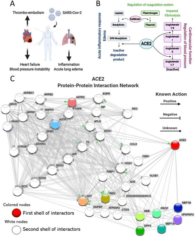
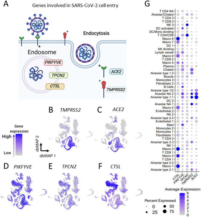
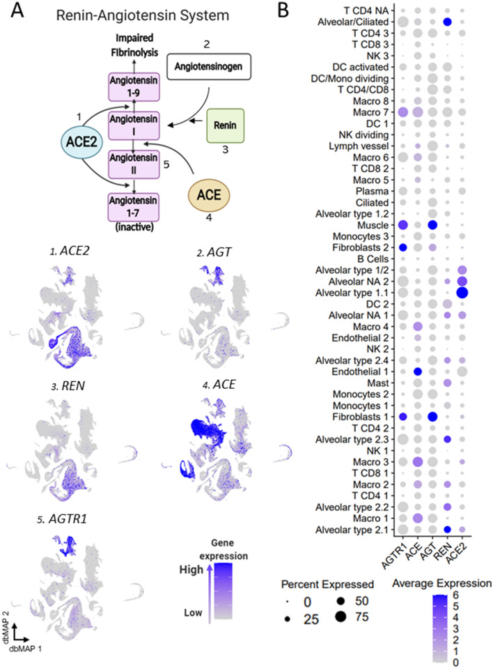
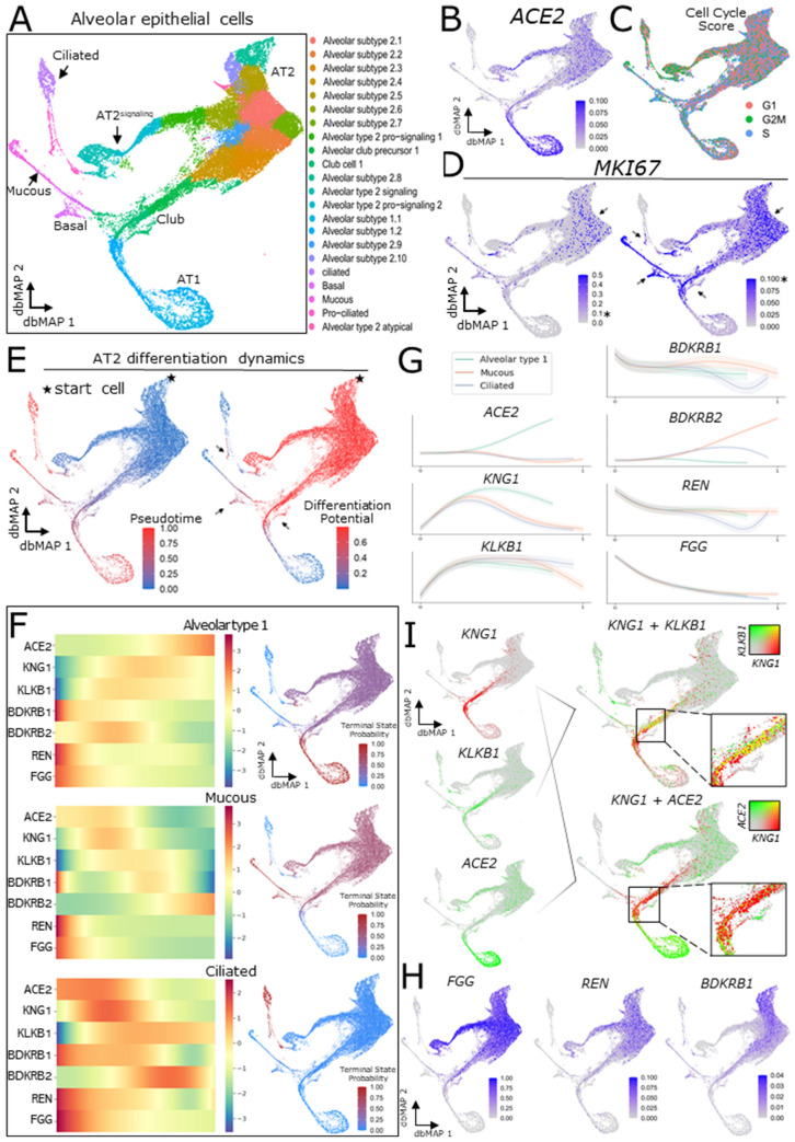
SARS-CoV-2, the pathogenic agent of COVID-19, employs angiotensin converting enzyme-2 (ACE2) as its cell entry receptor. Clinical data reveal that in severe COVID-19, SARS-CoV-2 infects the lung, leading to a frequently lethal triad of respiratory insufficiency, acute cardiovascular failure, and coagulopathy. Physiologically, ACE2 plays a role in the regulation of three systems that could potentially be involved in the pathogenesis of severe COVID-19: the kinin-kallikrein system, resulting in acute lung inflammatory edema; the renin-angiotensin system, promoting cardiovascular instability; and the coagulation system, leading to thromboembolism. Here we assembled a healthy human lung cell atlas meta-analysis with ~ 130,000 public single-cell transcriptomes and show that key elements of the bradykinin, angiotensin and coagulation systems are co-expressed with ACE2 in alveolar cells and associated with their differentiation dynamics, which could explain how changes in ACE2 promoted by SARS-CoV-2 cell entry result in the development of the three most severe clinical components of COVID-19.


Conflict of interest statement

The authors declare no competing interests.

Figures

Figure 1
Figure 1
COVID-19 clinical triad and ACE2 physiological roles link COVID-19 to ACE2 dysfunction. (A) Schematic representation of the clinical triad that play central role in severe COVID-19. (B) Schematic representation of the key components of the kinin–kallikrein, renin–angiotensin and coagulation systems and their interfaces with ACE2. (C) ACE2 Protein–Protein Interaction Network. ACE2 interactome was retrieved with the data-mining toolkit string-db (https://version-11-0.string-db.org/cgi/network.pl?taskId=KwL0Kho7pBZf). First shell interactors of ACE2 were set as colored nodes, while second shell interactors were set as white nodes. Edges indicate known molecular action of a protein node regarding another protein node. ACE2 is directly connected to components of the kinin–kallikrein (bradykinin), renin–angiotensin and coagulation systems.
Figure 2
Figure 2
Generation of an Integrated Lung Cell Atlas through integration of public data reveal fibroblasts and alveolar cells expressing ACE2 in the human lung. (a) Public data from single-cell RNA sequencing (scRNAseq) studies on the human lung was retrieved. Reyfman et al., Madissoon et al. and Human Cell Landscape control datasets were individually analyzed and batch-corrected with Seurat v3 anchor-based integration after filtering and normalization. Batch-corrected data representing each study was used to assemble an integrated dataset. This integrated dataset was annotated with the assistance of label transferred from Travaglini et al. data. (b) The Human Lung Integrated Cell Atlas represented in two-dimensional diffusion-based Manifold Approximation Embedding (dbMAP). dbMAP organizes the visualization to preserve as much of the original data structure as possible in a comprehensive way. In this representation, each cell is a point mapped to an embedding so that its x, y coordinates represent its relative transcriptional identity, i.e. its phenotypic signal. Cells are colored by their assigned cluster. (c) Visualization of ACE2 expression on the human lung dbMAP embedding. ACE2 is consistently expressed in alveolar cells and scattered in fibroblasts, being practically absent from other cell types. NK Natural Killer cell; DC Dendritic Cell; NA Not Assigned.
Figure 3
Figure 3
Lung expression of genes involved in SARS-Cov-2 cell entry is selective to alveolar cells. (a) Schematic representing SARS-Cov-2 cell entry mechanism. TMPRSS2 cleaves SARS-Cov-2 spike protein, allowing it to bind to ACE2, its functional receptor. It is known that other genes such as PIKFYVE, TPCN2 and CTSL also play a role in SARS-Cov-2 endocytosis and cellular contamination (b-f). Visualization of gene expression in dbMAP embeddings of the lung atlas. b. TMPRSS2 expression is restricted to alveolar cells. c. ACE2 expression is selective to alveolar cells and fibroblasts. (df) PIKFYVE, TPCN2 and CTSL are expressed consistently throughout the majority of lung cell types. CTSL expression in macrophages (g). Dot plot visualization of expression of genes involved in SARS-Cov-2 cell entry in human lung cell types. ACE2 expression is selective to alveolar clusters, similarly to TMPRSS2, but with the particularity of being lowly expressed in a large portion of endothelial cells. PIKFYVE, CTSL and TPCN2 are expressed by all clusters, but CTSL expression is much higher in macrophages, while TPCN2 expression is higher in NK and T CD4/CD8 clusters.
Figure 4
Figure 4
Gene expression of components of the kinin–kallikrein system point to alveolar cells role. (a) Schematic representing the main players and steps of the bradykinin system, also known as the kinin–kallikrein system. High molecular weight kininogen (KNG1) conversion into bradykinin is catalyzed by kallikrein. Bradykinin binds to the bradykinin receptor 2 (BDKRB2), but it can be converted into DR9-bradykinin, which activates the bradykinin receptor 1 (BDKRB1) until it is degraded by ACE2 into inactive degradation products. Bradykinin can also be directly converted into inactive degradation products by ACE. 16 Visualization of gene expression in dbMAP embeddings of the lung atlas. 1 KNG1 is selectively expressed in alveolar cells. 2 KLKB1 is expressed in endothelial and lymph vessel cells, and sparsely expressed in alveolar cells. 3 BDKRB2 expression is selective to endothelial cells. 4 ACE expression is selective to endothelial cells and macrophages. 5 BDKRB1 expression is selective to endothelial cells and fibroblasts, but also detected in low levels in alveolar cells. 6 ACE2 expression is restricted to alveolar cells. (b) Dot plot visualization of expression of genes involved in the bradykinin system in the lung atlas cell types. ACE2 and KNG1 gene expression is selective to alveolar clusters, despite ACE2 being lowly expressed in a large portion of endothelial cells and KNG1 also being lowly expressed in a large portion of clusters of macrophages. KLKB1 is highly expressed in endothelial and lymph vessel cells, but also lowly expressed in T CD4 clusters. BDKRB2 is highly expressed in endothelial and alveolar type 1.1 cells, and also less expressed by a large portion of B/plasma cells, and of macrophages clusters 7 and 8. ACE is selectively expressed in endothelial cells and in macrophages. BDKRB1 expression is selective to fibroblasts, although various clusters express it in low levels. ACE2 expression is restricted to alveolar cells.
Figure 5
Figure 5
Gene expression of components of the renin–angiotensin system point towards alveolar cells role. (a) Schematic representing the main players and steps of the angiotensin system, also known as the renin–angiotensin system (RAS). ACE2 catalyzes the conversion of angiotensin I to angiotensin 1–9, and of angiotensin II into inactive angiotensin 1–7. Renin catalyzes the conversion of angiotensinogen (AGT) into angiotensin I. ACE catalyzes the conversion of angiotensin I into angiotensin II, which binds to its receptor AGTR1. 15 Visualization of gene expression in dbMAP embeddings of the lung atlas. 1 ACE2 expression is restricted to alveolar cells. 2 AGT is expressed in fibroblasts and smooth muscle cells. 3 REN expression is restricted to alveolar cells. 4 ACE expression is selective to endothelial cells and macrophages. 5 AGTR1 expression is selective to fibroblasts and smooth muscle cells. (b) Dot plot visualization of the expression of genes involved in the angiotensin system in the lung atlas cell types. ACE2 expression is restricted to alveolar cell clusters, similarly to REN, which is virtually absent from remaining clusters. AGT is highly expressed in fibroblasts and muscle, albeit being lowly expressed in a large portion of cells from other clusters. ACE is preferentially expressed in endothelial cells and macrophage clusters. AGTR1 is highly expressed in smooth muscle cells and fibroblasts, and lowly expressed in macrophages and alveolar cells.
Figure 6
Figure 6
Gene expression of components of the coagulation system. (a) Schematic representing the final step of the coagulation system. Plasma kallikrein (KLKB1) catalyzes the conversion of plasminogen into plasmin. Plasminogen is activated by tissue plasminogen activator (PLAT), which is regulated by serpine 1 (SERPINE1). Plasminogen conversion into plasmin promote fibrinolysis by degradation of fibrin networks generated from fibrinogen (FGG) into inactive fragments. ACE2 plays a role in this process by catalyzing the conversion of angiotensin I to angiotensin 1–9, which inhibits fibrinolysis. 15 Visualization of gene expression in dbMAP embeddings of the lung atlas. 1 KLKB1 is expressed in endothelial cells and sparsely in alveolar cells. 2 SERPINE1 is highly expressed in fibroblasts and in endothelial and smooth muscle cells, and present in lower levels in macrophages. 3 PLAT is mostly expressed in endothelial cells and fibroblasts. 4 FGG expression is restricted to alveolar cells. 5 ACE2 expression is restricted to alveolar cell clusters. (b) Dot plot visualization of the expression of genes involved in the coagulation system in the lung atlas cell types. ACE2 expression is restricted to alveolar cell clusters. PLAT expression is higher in endothelial cells, lymph vessel cells and mast cells. FGG expression is restricted to alveolar cell clusters, being higher in alveolar type 2.1 and type 2.2, alveolar/ciliated and alveolar NA clusters.
Figure 7
Figure 7
ACE2 and components of the KKS, RAS and CS are differentially expressed throughout alveolar epithelium differentiation dynamics. (a) Two-dimensional dbMAP embedding of 37,449 alveolar cells transcriptomes. Cells are colored by their assigned subtype. Main cell types are indicated. (b) Visualization of ACE2 expression in the dbMAP embedding. The expression pattern prioritizes AT1 cells, and less so AT2 cells. (c) Cell cycle scoring of each single-cell and its assigned cell-phase. Note how all phases are detected across the embedding, albeit at different proportions. (d) MKI67 expression visualization in the dbMAP embedding with two different coloring thresholds. AT2 cells express MKI67 at higher levels (left; arrow), and mucous, basal, club and pro-ciliated cells present lower, yet consistent levels of MKI67 expression (right; arrows). The * indicates a fixed expression level, for easier visualization. (e) AT2 progeny differentiation dynamics. The star represents the start cell used in computations. Left: pseudotime score for each cell, i.e. how much each cell is advanced in the differentiation trajectory. Right: differentiation potential for each cell, i.e. the probability that cell i transit into an adjacent cell j, also seen as entropy. Note that the intermediate states (pointed by arrows) present a higher differentiation potential than terminally differentiated cells. (f) The three terminal states detected for the AT2 progeny—alveolar type 1 (AT1), mucous and ciliated cells. The heatmaps on the left column indicate relative expression changes (left to right) throughout the differentiation trajectories shown on the right column that correspond to terminal states. (g) Gene expression trends of ACE2 and components of the KKS, RAS and CS expressed in alveolar cells. ACE2 expression is directly associated with the maturation of AT1 cells, whereas KNG1 and KLKB1 are preferentially expressed in intermediate states. REN, FGG and BDKRB1 are preferentially expressed in AT2 progenitors at the beginning of the differentiation process, and BDKRB2 expression is directly associated with the differentiation mucous cells. (i) Visualization of co-expression (yellow) by coloring overlap. KNG1 (red) is co-expressed with KLKB1 (green, top) in intermediate states corresponding to club cells, but barely co-expressed with ACE2 (green, bottom). (h) Visualization of FGG, REN and BDKRB1 expression in the dbMAP embedding. As shown in (g), these genes are preferentially expressed by AT2 progenitors, being undetected in terminally differentiated states.
Figure 8
Figure 8
A theoretical model for Sars-Cov-2 pathogenesis. (a) Schematic representation of the identified alveolar epithelium progeny, with AT2 precursors giving rise to club and AT2-signaling cells. The latter then gives rise to pro-ciliated and ciliated cells, whereas the latter give rise to basal, mucous and AT1 precursor cells. AT1 precursors which are ACE2-/KNG1+ /KLKB1+ give rise to mature AT1 cells, which highly express ACE2 during their differentiation process. Selective disruption of the alveolar niche turnover dynamics could thus be implicated in Sars-Cov-2 pathogenesis, and we encourage further studies investigating these associations. (b) Schematic representation of the healthy alveolus prior to Sars-Cov-2 binding to ACE2 (1). Either by structural ACE2 changes or by disruption of the ACE2+ population, Sars-Cov-2 infection potentially interferes with two key ACE2 roles: (2a) converting Ang II to Ang 1–7, thus compromising alveolar cell survival signaling; and (2b) degradation of bradykinin into inactive products. Pro-AT1 (club) cells highly and selectively express kininogen and kallikrein (3), which jointly leads to the production of bradykinin. Kallikrein is also involved in converting plasminogen into plasmin (not shown). Infection derived disruption of the alveolar niche with selective preservation of ACE2-/KNG1+ /KLKB1+ pro-AT1 club cells could be associated with disturbance of the KKS, leading to severe inflammation and altered coagulation.

References

    1. Berlin DA, Gulick RM, Martinez FJ. Severe covid-19. N. Engl. J. Med. 2020 doi: 10.1056/NEJMcp2009575. - DOI - PubMed
    1. Weiss P, Murdoch DR. Clinical course and mortality risk of severe COVID-19. Lancet. 2020;395:1014–1015. - PMC - PubMed
    1. Zhou F, et al. Clinical course and risk factors for mortality of adult inpatients with COVID-19 in Wuhan, China: a retrospective cohort study. Lancet. 2020;395:1054–1062. - PMC - PubMed
    1. Richardson S, et al. Presenting characteristics, comorbidities, and outcomes among 5700 patients hospitalized with Covid-19 in the New York City area. JAMA. 2020 doi: 10.1001/jama.2020.6775. - DOI - PMC - PubMed
    1. Guan WJ, et al. Clinical characteristics of coronavirus disease 2019 in China. N. Engl. J. Med. 2020;382:1708–1720. - PMC - PubMed

Publication types

MeSH terms