. 2020 Nov 3:13:11211-11220.
doi: 10.2147/OTT.S267937. eCollection 2020.

LncRNA XIST Inhibits the Progression of Oral Squamous Cell Carcinoma via Sponging miR-455-3p/BTG2 Axis

Affiliations

LncRNA XIST Inhibits the Progression of Oral Squamous Cell Carcinoma via Sponging miR-455-3p/BTG2 Axis

Qingbin Li et al. Onco Targets Ther. .

Abstract

Objective: Oral squamous cell carcinoma (OSCC) is one of the most common cancers, accounting for over 90% of malignant lesions in the oral cavity. Long non-coding RNAs play an important role in the development of OSCC. This study aimed to investigate the effects of lncRNA XIST on the malignant behaviors of OSCC cells and its possible molecular mechanisms.

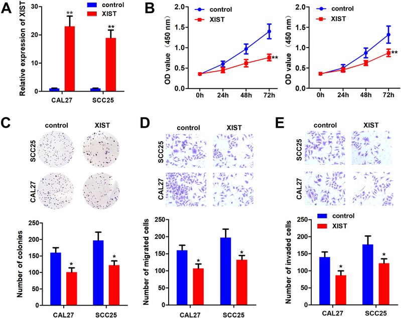
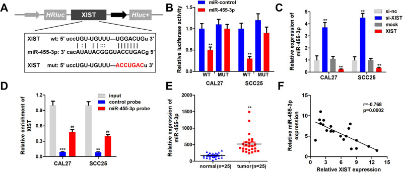
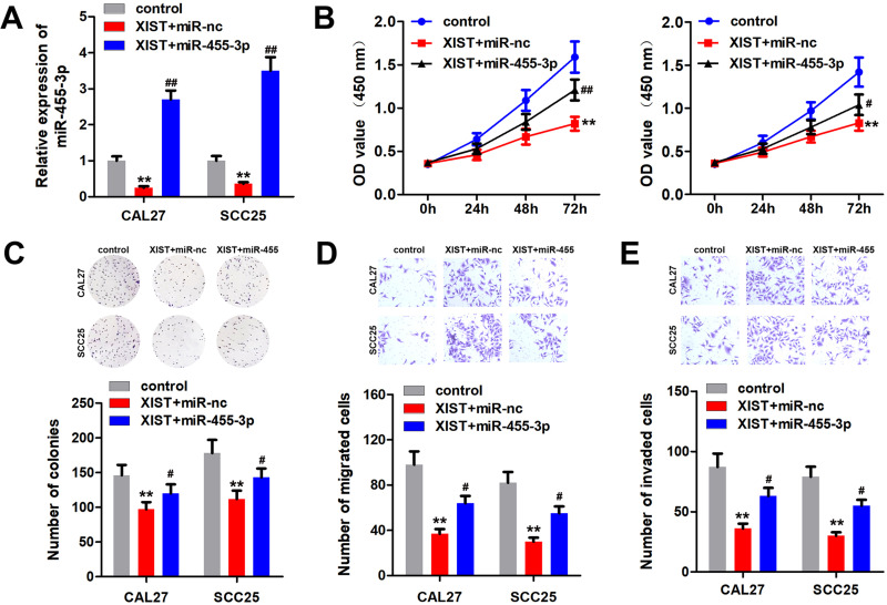
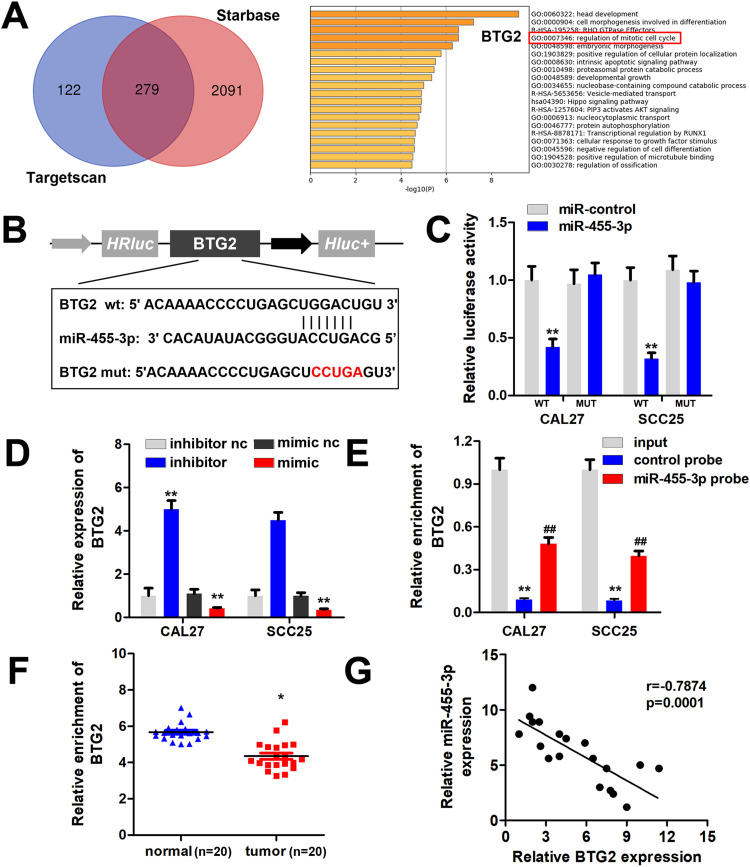
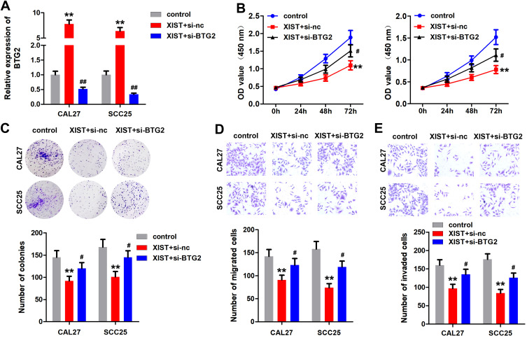
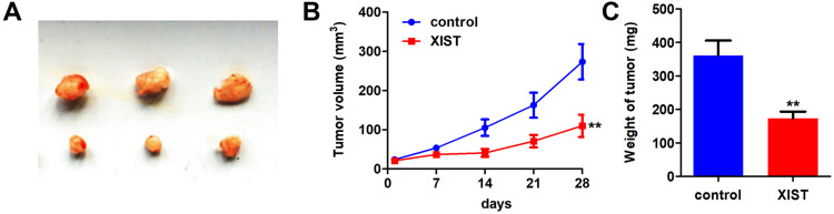
Methods: Real-time quantitative PCR and Western blot were used to detect the RNA and protein level, respectively. CAL27 and SCC25 cells with the lowest expression level of XIST were used for further study. MTT, transwell assay, colony formation, and xenograft model were applied to examine the effect of XIST on the progression of OSCC. FISH assay was performed to investigate the co-location of XIST and miR-455-3p in OSCC cells. The bioinformatics analysis, luciferase, and RNA pull down assay were utilized to predict and verify the target genes of miR-455-3p.

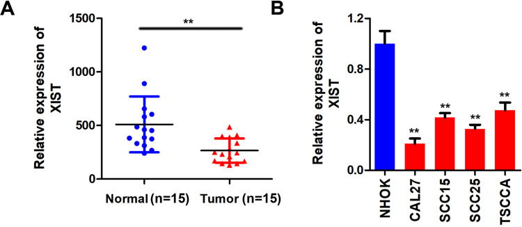
Results: XIST was downregulated in OSCC tissues and cell lines. Overexpression of XIST inhibited the proliferation, migration, and invasion ability of OSCC cells. Bioinformatics analysis and luciferase reporter assay confirmed XIST could bind to miR-455-3p. Besides, miR-455-3p directly targeted BTG2 in OSCC cells. Rescue experiments further confirmed the positive interaction between miR-455-3p and XIST as well as between miR-455-3p and BTG2.

Conclusion: XIST was down-regulated in OSCC. XIST regulated the expression of BTG2 via sponging miR-455-3p. XIST/miR-455-3p/BTG2 signal axis inhibited the malignant progression of OSCC.

Keywords: BTG2; OSCC; XIST; miR-455-3p.


Conflict of interest statement

The authors report no conflicts of interest in this work.

Figures

Figure 1
Figure 1
XIST is lowly expressed in OSCC. (A and B) qPCR was used to detect the expression of XIST in the OSCC tissues and cell lines compare to the normal tissues and oral mucosa epithelium cells. **P<0.01.
Figure 2
Figure 2
XIST inhibited the malignant behavior of OSCC. pcDNA3.1/XIST was transfected to OSCC cells to overexpressing XIST. (A) qPCR was used to assess the XIST expression. (B and C) MTT and colony formation assay were performed to detect the cell proliferation of OSCC cells after overexpression of XIST. (D and E) Transwell assay was used to evaluate the migration and invasion of OSCC cells. *P<0.05, **P<0.01.
Figure 3
Figure 3
XIST sponges miR-455-3p in OSCC cells. (A) Bioinformatics analysis showed the binding site between XIST and miR-455-3p. (B) Luciferase assay was carried out to verify the combination of XIST and miR-455-3p. (C) qPCR was used to assess the expression of miR-455-3p. (D) RNA pulled down experiments was performed to confirm the interaction between XIST and miR-455-3p. (E) qPCR was used to detect the expression of miR-455-3p in OSCC tissues. (F) Pearson analysis was performed to evaluate the correlation between XIST and miR-455-3p. **P<0.01, ***P<0.001, ##P<0.01.
Figure 4
Figure 4
miR-455-3p reversed the effect of XIST in OSCC cells. OSCC cells were transfected with pcDNA3.1/XIST, miR-455-3p mimic or their combination. (A) miR-455-3p expression was detected by qPCR. (B and C) MTT and colony formation assay were performed to detect the cell proliferation of OSCC cells. (D and E) Transwell assay was used to evaluate the migration and invasion of OSCC cells. **P<0.01, #P<0.05, ##P<0.01.
Figure 5
Figure 5
miR-455-3p targets BTG2 in OSCC cells. (A) Bioinformatics analysis based on the TargetScan and Starbase database was performed to screen the targets of miR-455-3p. Enrichment analysis of these 279 potential target genes was performed using Metaspace software. (B) The binding site between miR-455-3p and BTG2 was showed. (C) Luciferase assay was carried out to verify the combination of miR-455-3p and BTG2. (D) qPCR was used to assess the BTG2 expression in each group. (E) RNA pulled down experiments was performed to confirm the interaction between miR-455-3p and BTG2. (F) qPCR was used to assess the expression level BTG2 in OSCC tissues. (G) Pearson analysis was performed to evaluate the correlation between miR-455-3p and BTG2 expression level. *P<0.05, **P<0.01, ##P<0.01.
Figure 6
Figure 6
Knockdown of BTG2 reverses the anti-cancer effect of XIST. OSCC cells were transfected with pcDNA3.1/XIST, si-BTG2 or their combination. (A) The expression of BTG2 was detected by qPCR. (B and C) MTT and colony formation assay were performed to detect the cell proliferation of OSCC cells. (D and E) Transwell assay was used to evaluate the migration and invasion of OSCC cells. **P<0.01, #P<0.05, ##P<0.01.
Figure 7
Figure 7
XIST inhibited tumor proliferation of OSCC. (A) CAL27 cell line stably expressing pcDNA3.1/XIST was used to establish the Xenografts mice model. (A) The image of the tumors in each group. (B) The growth curve of the tumors was made. (C) The wight of tumor in each group was detected. **P<0.01.

References

    1. Momen-Heravi F, Bala S. Emerging role of non-coding RNA in oral cancer. Cell Signal. 2018;42:134–143. doi:10.1016/j.cellsig.2017.10.009 - DOI - PubMed
    1. Yang Y, Chen D, Liu H, Yang K. Increased expression of lncRNA CASC9 promotes tumor progression by suppressing autophagy-mediated cell apoptosis via the AKT/mTOR pathway in oral squamous cell carcinoma. Cell Death Dis. 2019;10(2):41. doi:10.1038/s41419-018-1280-8 - DOI - PMC - PubMed
    1. Hazawa M, Lin DC, Handral H, et al. ZNF750 is a lineage-specific tumour suppressor in squamous cell carcinoma. Oncogene. 2017;36(16):2243–2254. doi:10.1038/onc.2016.377 - DOI - PMC - PubMed
    1. Wang Y, Zhang X, Wang Z, et al. LncRNA-p23154 promotes the invasion-metastasis potential of oral squamous cell carcinoma by regulating glut1-mediated glycolysis. Cancer Lett. 2018;434:172–183. doi:10.1016/j.canlet.2018.07.016 - DOI - PubMed
    1. Peng WX, Koirala P, Mo YY. LncRNA-mediated regulation of cell signaling in cancer. Oncogene. 2017;36(41):5661–5667. doi:10.1038/onc.2017.184 - DOI - PMC - PubMed