

Ion Channel Signature in Healthy Pancreas and Pancreatic Ductal Adenocarcinoma
- PMID: 33178018
- PMCID: PMC7596276
- DOI: 10.3389/fphar.2020.568993
Ion Channel Signature in Healthy Pancreas and Pancreatic Ductal Adenocarcinoma
Abstract
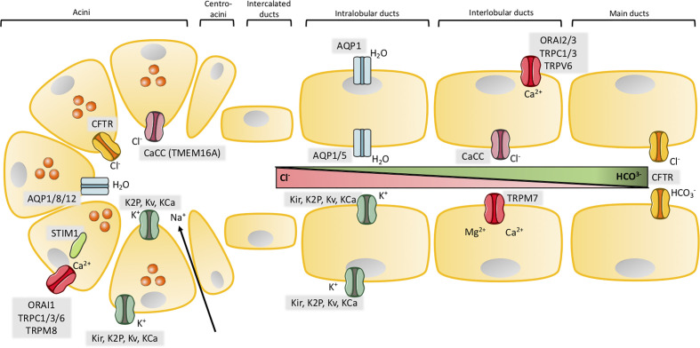
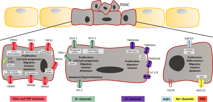
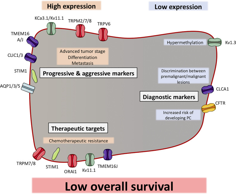
Pancreatic ductal adenocarcinoma (PDAC) is the fourth most common cause of cancer-related deaths in United States and Europe. It is predicted that PDAC will become the second leading cause of cancer-related deaths during the next decades. The development of PDAC is not well understood, however, studies have shown that dysregulated exocrine pancreatic fluid secretion can contribute to pathologies of exocrine pancreas, including PDAC. The major roles of healthy exocrine pancreatic tissue are secretion of enzymes and bicarbonate rich fluid, where ion channels participate to fine-tune these biological processes. It is well known that ion channels located in the plasma membrane regulate multiple cellular functions and are involved in the communication between extracellular events and intracellular signaling pathways and can function as signal transducers themselves. Hereby, they contribute to maintain resting membrane potential, electrical signaling in excitable cells, and ion homeostasis. Despite their contribution to basic cellular processes, ion channels are also involved in the malignant transformation from a normal to a malignant phenotype. Aberrant expression and activity of ion channels have an impact on essentially all hallmarks of cancer defined as; uncontrolled proliferation, evasion of apoptosis, sustained angiogenesis and promotion of invasion and migration. Research indicates that certain ion channels are involved in the aberrant tumor growth and metastatic processes of PDAC. The purpose of this review is to summarize the important expression, localization, and function of ion channels in normal exocrine pancreatic tissue and how they are involved in PDAC progression and development. As ion channels are suggested to be potential targets of treatment they are furthermore suggested to be biomarkers of different cancers. Therefore, we describe the importance of ion channels in PDAC as markers of diagnosis and clinical factors.
Keywords: biomarkers; exocrine pancreas; ion channels; pancreatic ductal adenocarcinoma; signaling pathways.
Copyright © 2020 Schnipper, Dhennin-Duthille, Ahidouch and Ouadid-Ahidouch.
Figures
Similar articles
-
Ion Channels Orchestrate Pancreatic Ductal Adenocarcinoma Progression and Therapy.Front Pharmacol. 2021 Jan 19;11:586599. doi: 10.3389/fphar.2020.586599. eCollection 2020. Front Pharmacol. 2021. PMID: 33841132 Free PMC article. Review.
-
Transportome Malfunctions and the Hallmarks of Pancreatic Cancer.Rev Physiol Biochem Pharmacol. 2021;181:105-127. doi: 10.1007/112_2020_20. Rev Physiol Biochem Pharmacol. 2021. PMID: 32770395 Review.
-
Integrated expression profiling of potassium channels identifys KCNN4 as a prognostic biomarker of pancreatic cancer.Biochem Biophys Res Commun. 2017 Dec 9;494(1-2):113-119. doi: 10.1016/j.bbrc.2017.10.072. Epub 2017 Oct 16. Biochem Biophys Res Commun. 2017. PMID: 29050937
-
Ca2+ Signaling and Its Potential Targeting in Pancreatic Ductal Carcinoma.Cancers (Basel). 2021 Jun 21;13(12):3085. doi: 10.3390/cancers13123085. Cancers (Basel). 2021. PMID: 34205590 Free PMC article. Review.
-
Role of ion channels in gastrointestinal cancer.World J Gastroenterol. 2019 Oct 14;25(38):5732-5772. doi: 10.3748/wjg.v25.i38.5732. World J Gastroenterol. 2019. PMID: 31636470 Free PMC article. Review.
Cited by
-
Whole genome sequencing identifies rare germline variants enriched in cancer related genes in first degree relatives of familial pancreatic cancer patients.Clin Genet. 2021 Nov;100(5):551-562. doi: 10.1111/cge.14038. Epub 2021 Aug 3. Clin Genet. 2021. PMID: 34313325 Free PMC article.
-
Survival-Associated Cellular Response Maintained in Pancreatic Ductal Adenocarcinoma (PDAC) Switched Between Soft and Stiff 3D Microgel Culture.ACS Biomater Sci Eng. 2024 Apr 8;10(4):2177-2187. doi: 10.1021/acsbiomaterials.3c01079. Epub 2024 Mar 11. ACS Biomater Sci Eng. 2024. PMID: 38466617 Free PMC article.
-
Examining the effects of biofield therapy through simultaneous assessment of electrophysiological and cellular outcomes.Sci Rep. 2024 Dec 2;14(1):29221. doi: 10.1038/s41598-024-79617-3. Sci Rep. 2024. PMID: 39622875 Free PMC article.
-
Synergistic effects of agonists and two-pore-domain potassium channels on secretory responses of human pancreatic duct cells Capan-1.Pflugers Arch. 2023 Mar;475(3):361-379. doi: 10.1007/s00424-022-02782-9. Epub 2022 Dec 19. Pflugers Arch. 2023. PMID: 36534232 Free PMC article.
-
Molecular mechanisms of pain in acute pancreatitis: recent basic research advances and therapeutic implications.Front Mol Neurosci. 2023 Dec 22;16:1331438. doi: 10.3389/fnmol.2023.1331438. eCollection 2023. Front Mol Neurosci. 2023. PMID: 38188196 Free PMC article. Review.
References
Publication types
LinkOut - more resources
Full Text Sources
Research Materials