. 2020 Oct 15:11:555245.
doi: 10.3389/fimmu.2020.555245. eCollection 2020.

Non-Canonical WNT5A Signaling Through RYK Contributes to Aggressive Phenotype of the Rheumatoid Fibroblast-Like Synoviocytes

Affiliations

Non-Canonical WNT5A Signaling Through RYK Contributes to Aggressive Phenotype of the Rheumatoid Fibroblast-Like Synoviocytes

Angela Rodriguez-Trillo et al. Front Immunol. .

Abstract

We hypothesized that WNT5A could contribute to the enhanced migration and invasiveness of rheumatoid arthritis fibroblast-like synoviocytes (RA FLS), which is one of the incompletely understood aspects of the RA FLS aggressive phenotype. This hypothesis is based on the previous evidence of a WNT5A role in both, RA and cell migration. Migration and invasion of RA FLS were assessed after incubation with recombinant Wnt5a (rWnt5a) or silencing of the endogenous WNT5A expression. The expression of WNT5A, WNT receptors, cytokines, chemokines, and metalloproteinases was quantified with RT-PCR. The WNT pathway was explored with gene silencing, antibody and pharmacological inhibition followed by migration assays and phosphoprotein western blots. Here, we reported that rWnt5a promoted migration and invasion of RA FLS, whereas knockdown of the endogenous WNT5A reduced them. These effects were specific to the RA FLS since they were not observed in FLS from osteoarthritis (OA) patients. Also, rWnt5a induced the expression of IL6, IL8, CCL2, CXCL5, MMP1, MMP3, MMP9, and MMP13 from baseline or potentiating the TNF induction, WNT5A signaling required the RYK receptor and was mediated through the WNT/Ca2+ and the ROCK pathway. These pathways involved the RYK and ROCK dependent activation of the p38, ERK, AKT, and GSK3β kinases, but not the activation of JNK. Together these findings indicate that WNT5A contributes to the enhanced migration and invasiveness of RA FLS through RYK and the specific activation of ROCK and downstream kinases.

Keywords: MAPK; RYK; WNT5A; fibroblast-like synoviocytes; inflammatory response; invasion; migration; rheumatoid arthritis.


Figures

Figure 1
Figure 1
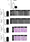
The migration and invasion of RA FLS are promoted by WNT5A. (A) Expression of WNT5A mRNA relative to that for β-actin in RA and OA patients was determined. (B, C) The FLS from RA (B) and OA (C) patients were stimulated with 400 ng/ml rWnt5a and the migration rate as percentage of the control was measured by wound-healing assays at 96 h (D, E) Percentage of RA FLS (D) and OA FLS (E) stimulated or not with 400 ng/ml rWnt5a, invading the Matrigel coated inserts at 48 h compared with the controls without rWnt5a. Values are the Mean ± Standard error of the mean (SEM) of FLS from 5 to 8 RA and OA patients obtained from a total of 9 and 8 independent experiments in migration and invasion assays, respectively. *P < 0.05, by Mann Whitney U test (A) and Wilcoxon matched-pairs test (B–E).
Figure 2
Figure 2
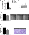
WNT5A suppression reduces migration and invasion in RA FLS. (A, B). FLS were transfected with WNT5A or control siRNA and the efficiency of silencing was determined by real-time PCR (A) and western blot (B). A representative blot is shown. (C, D) Analysis of migration (C) and invasion (D) in RA FLS transfected with WNT5A or control siRNA. Representative images are shown. Values are the Mean ± Standard error of the mean (SEM) of FLS from 6 patients obtained from 5 independent experiments in each assay. * and # indicates P < 0.05, by Wilcoxon matched-pairs test.
Figure 3
Figure 3
The gene expression of inflammatory mediators and metalloproteinases in RA FLS are promoted by WNT5A in RA FLS. (A, B) Fold change of IL6, IL8, CCL2, CCL5, CXCL5, MMP1, MMP2, MMP3, MMP9, and MMP13 mRNA expression in RA FLS stimulated with 400 ng/ml rWnt5a alone or in combination with 10 ng/ml TNF (A), or by transfection with WNT5A or control siRNA (CCL5 was not included) (B). Values are the Mean ± Standard error of the mean (SEM) of FLS from 7 patients with RA obtained from 7 independent experiments. * and # indicates P < 0.05, by Wilcoxon matched-pairs test.
Figure 4
Figure 4
Identification of the receptors involved in WNT5A signaling in RA FLS. (A) Expression of the WNT receptors in FLS from 6 RA patients as assessed by real-time PCR. (B) Effect of silencing RA FLS FZD1, FZD2, FZD4, FZD7 and ROR1 by siRNA transfection on the RA FLS migration stimulated with 400 ng/ml rWnt5a in comparison with the siRNA control transfection as measured by wound-healing assays at 96 h. (C, D) Effect of the anti ROR2 antibody (α-ROR2), 4 μg/ml (C), or the anti-RYK antibody (α-RYK), 1 μg/ml (D) on the basal and rWnt5a-induced migration (400 ng/ml) of RA FLS. Migration of RA FLS without treatment was the 100% (C, D). Values are the Mean ± Standard error of the mean (SEM) of FLS from 6 to 8 patients with RA obtained from four to seven independent experiments. * and # indicate P < 0.05, by Wilcoxon matched-pairs test.
Figure 5
Figure 5
Identification of the pathways involved in WNT5A signaling in RA FLS. (A) Effect of the BAPTA Ca2+ chelator, 10 μM on the basal and rWnt5a-induced migration (400 ng/ml) of RA FLS. (B) Representative blot of the time-course analysis of the changes in JNK phosphorylation induced by incubation with 400 ng/ml rWnt5a in comparison with 10 ng/ml TNF as determined by western blot. (C) Representative blot of the activation status of the JNK MAPK in RA FLS treated with the indicated doses of rWnt5a for 1 h in comparison with 10 ng/ml TNF. (D) Impact of the Y-27632 ROCK inhibitor (20 μM) on the rWnt5a-induced migration of the RA FLS. Migration of RA FLS without treatment was the 100% (A, C). Values are the Mean ± Standard error of the mean (SEM) of FLS from six to eight patients with RA obtained from 6 independent experiments. * and # indicate P < 0.05, by Wilcoxon matched-pairs test.
Figure 6
Figure 6
Downstream kinases activated by rWnt5a promote migration in RA FLS. (A) Analysis of the activation of p38, ERK MAPK, PI3K/AKT, and GSK3β by western blot in RA FLS treated with 400 ng/ml rWnt5a. (B) Effect of the MAPK-p38 inhibitor (SB 203580), PI3K inhibitor (LY 294002) or MAPK-ERK inhibitor (PD 98059) on the basal and rWnt5a-induced migration in RA FLS at 96 h. Values are the Mean ± Standard error of the mean (SEM) of FLS from six to eight patients with RA obtained from three to four independent experiments. * and # indicate P < 0.05, by Wilcoxon matched-pairs test.
Figure 7
Figure 7
Contribution of RYK/ROCK pathway to the activation of p38, ERK MAPK, PI3K/AKT, and GSK3β kinases. (A) Representative western blot showing the effect of the anti-RYK antibody (α-RYK) on the rWnt5a-induced activation of p38, ERK MAPK, PI3K/AKT, and GSK3β in RA FLS. (B) Effect of the Y-27632 ROCK inhibitor (20 μM) on the rWnt5a-induced activation of p38, ERK MAPK, PI3K/AKT, and GSK3β kinases determined by Western blot in RA FLS. Values are the Mean ± Standard error of the mean (SEM) of FLS from six to eight patients with RA obtained from three to four independent experiments. * and # indicate P < 0.05, by Wilcoxon matched-pairs test.
Figure 8
Figure 8
Schematic representation of the WNT5A signaling pathways involved in the migration of RA FLS. The participating molecules are shown together with the reagent used to demonstrate their respective involvement.

References

    1. Smolen JS, Aletaha D, Barton A, Burmester GR, Emery P, Firestein GS, et al. Rheumatoid arthritis. Nat Rev Dis Primers (2018) 4:18001. 10.1038/nrdp.2018.1 - DOI - PubMed
    1. Smolen JS, Aletaha D, McInnes IB. Rheumatoid arthritis. Lancet (2016) 388(10055):2023–38. 10.1016/S0140-6736(16)30173-8 - DOI - PubMed
    1. Niedermeier M, Pap T, Adelheid K. Therapeutic opportunities in fibroblasts in inflammatory arthritis. Best Pract Res Clin Rheumatol (2010) 24:527–40. 10.1016/j.berh.2010.02.002 - DOI - PubMed
    1. Pap T, Nawrath M, Heinrich J, Bosse M, Baier A, Hummel KM, et al. Cooperation of Ras- and c-Myc-dependent pathways in regulating the growth and invasiveness of synovial fibroblasts in rheumatoid arthritis. Arthritis Rheum (2004) 50:2794–802. 10.1002/art.20461 - DOI - PubMed
    1. Baier A, Meineckel I, Gay S, Pap T. Apoptosis in rheumatoid arthritis. Curr Opin Rheumatol (2003) 15:274–79. 10.1097/00002281-200305000-00015 - DOI - PubMed

Publication types

MeSH terms