. 2020 Oct 15:11:571334.
doi: 10.3389/fimmu.2020.571334. eCollection 2020.

Cutibacterium acnes Infection Induces Type I Interferon Synthesis Through the cGAS-STING Pathway

Affiliations

Cutibacterium acnes Infection Induces Type I Interferon Synthesis Through the cGAS-STING Pathway

Katrin Fischer et al. Front Immunol. .

Abstract

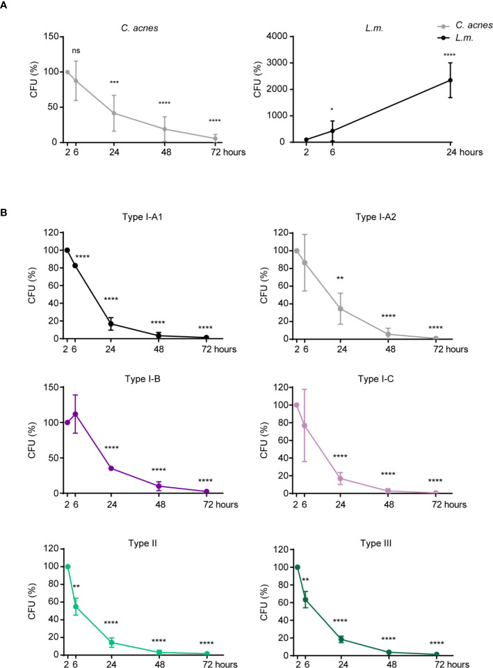
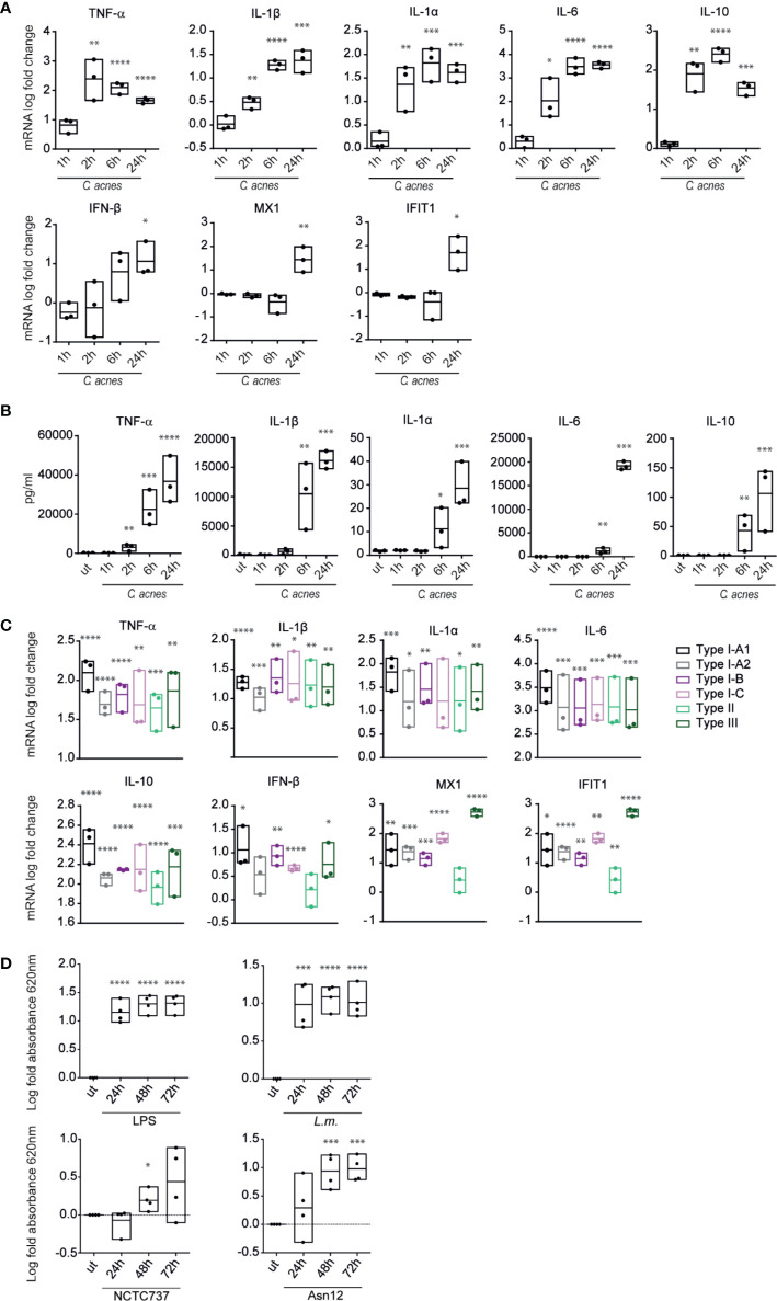
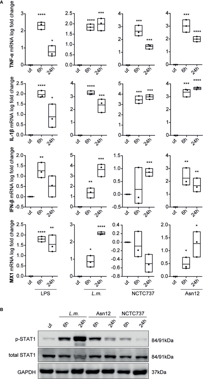
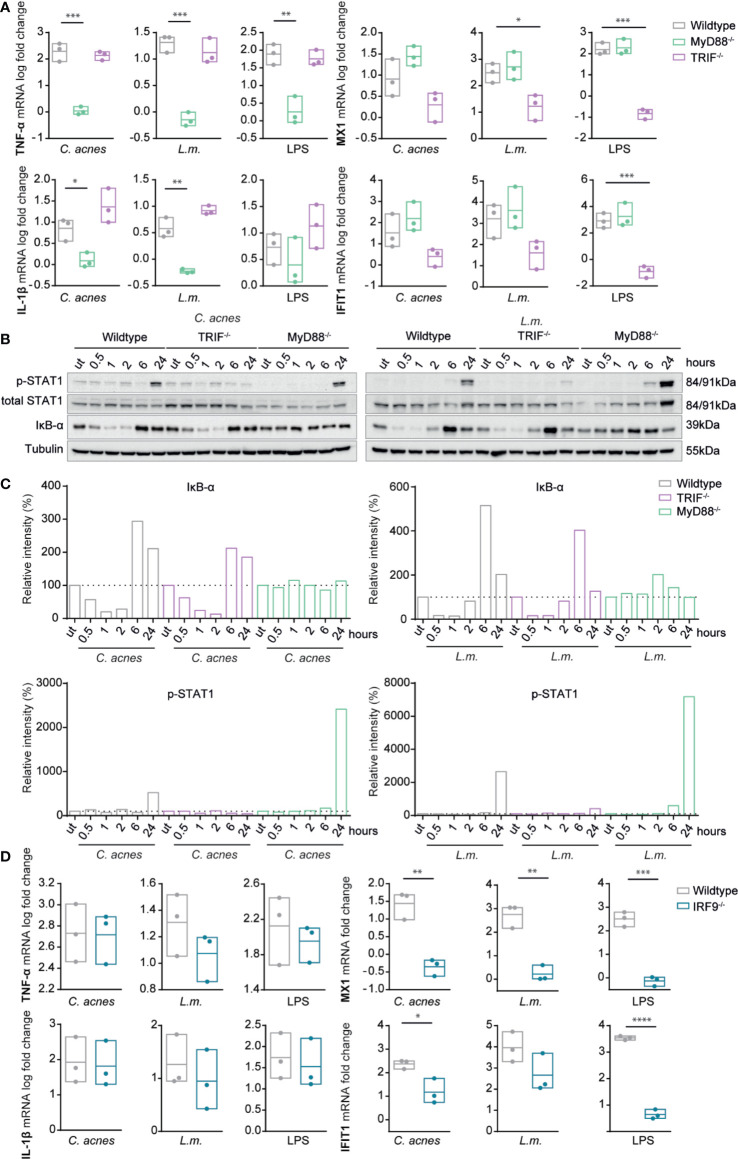
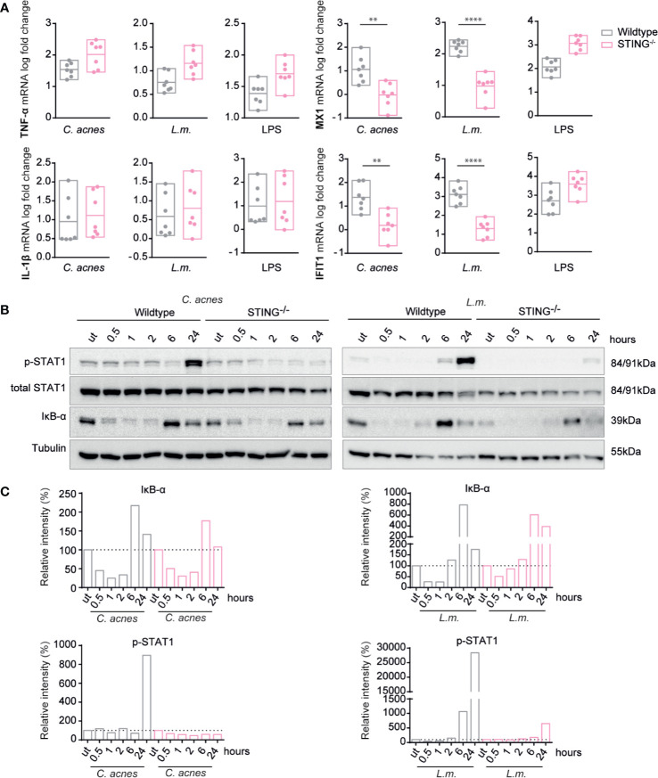
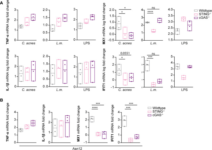
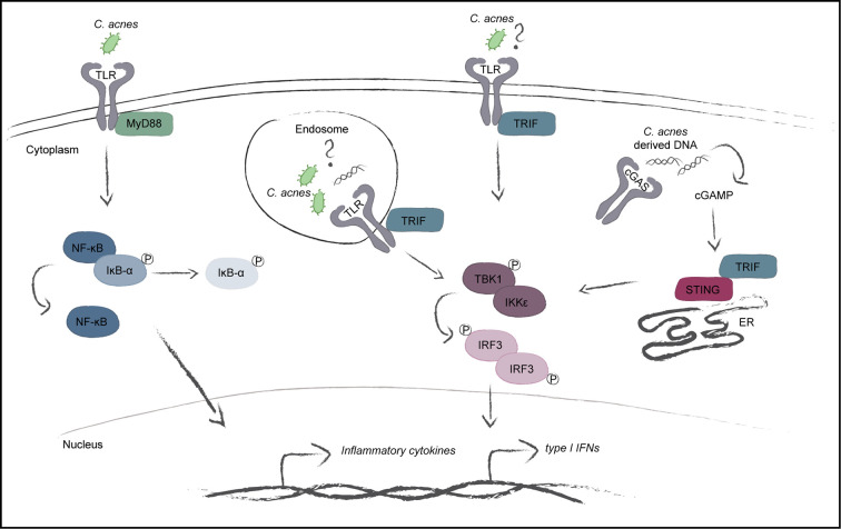
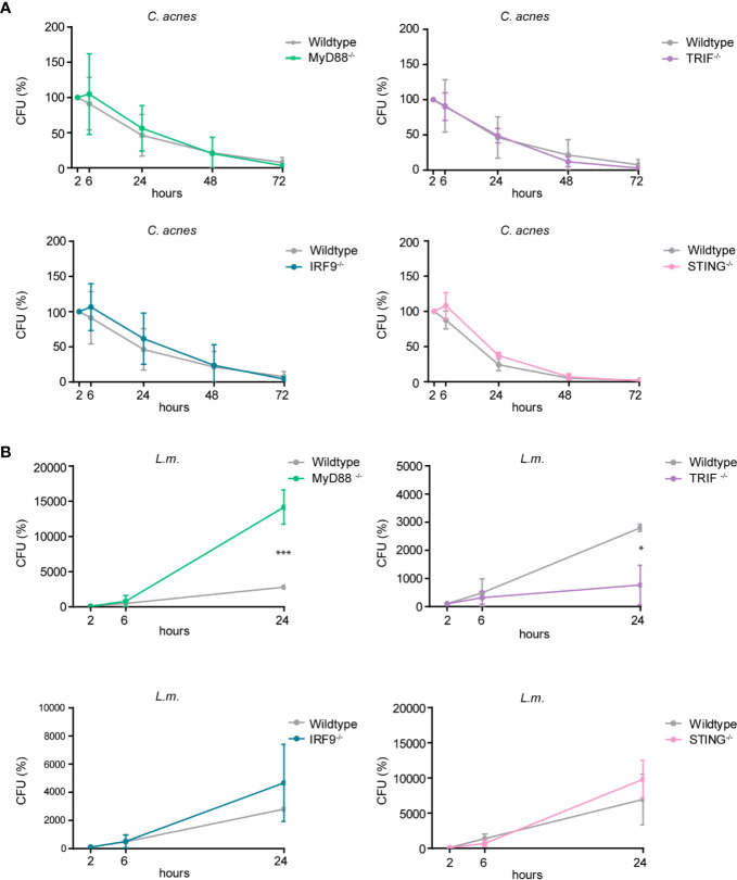
Cutibacterium (previously Propionibacterium) acnes is an anaerobic, Gram-positive commensal of the human body. The bacterium has been associated with a variety of diseases, including acne vulgaris, prosthetic joint infections, prostate cancer, and sarcoidosis. The accumulation of C. acnes in diseases such as acne and prostate cancer has been shown to correlate with enhanced inflammation. While the C. acnes-induced proinflammatory axis, via NF-κB and MAPK signaling and inflammasome activation, has been investigated over the last few decades, the potential role of C. acnes in triggering the type I interferon (IFN-I) pathway has not been addressed. Our results show that C. acnes induces the IFN-I signaling axis in human macrophages by triggering the cGAS-STING pathway. In addition, IFN-I signaling induced by C. acnes strongly depends on the adapter protein TRIF in a non-canonical manner; these signaling events occurred in the absence of any detectable intracellular replication of the bacterium. Collectively, our results provide important insight into C. acnes-induced intracellular signaling cascades in human macrophages and suggest IFN-I as a factor in the etiology of C. acnes-induced diseases. This knowledge may be valuable for developing novel therapies targeting C. acnes in diseases where the accumulation of the bacterium leads to an inflammatory pathology.

Keywords: Cutibacterium acnes; Listeria (L.) monocytogenes; NFkB = nuclear factor kappa b; STAT (signal transducer and activator of transcription); TIR-domain-containing adapter-inducing interferon-β; cyclic GMP-AMP synthase; interferon; stimulator of interferon genes.


Figures

Figure 1
Figure 1
Intracellular degradation of C. acnes by human macrophages. A CFU assay was performed using PMA-differentiated THP-1 cells. (A) THP-1 cells were either infected with C. acnes strain NCTC737 or L.m. for indicated timepoints. Graphs show the standard deviation and mean of five independent experiments. (B) Differentiated THP-1 cells were infected with one strain of each C. acnes phylotype: NCTC737 (phylotype IA1), P. acn31 (phylotype IA2), KPA171202 (phylotype IB), PV66 (phylotype IC), ATCC11828 (phylotype II), and Asn12 (phylotype III) for indicated timepoints. Graphs show the standard deviation and mean of three independent experiments. (A, B) Unpaired t-test of log transformed values compared to the 2-h timepoint was calculated for each timepoint. P-values (*P ≤ 0.05; **P ≤ 0.01; ***P ≤ 0.001; ****P ≤ 0.0001; ns, not significant).
Figure 2
Figure 2
Cytokine expression and secretion in response to C. acnes. (A) Differentiated THP-1 cells were infected with C. acnes strain NCTC737 for indicated timepoints. HPRT-normalized gene expression was measured using RT-qPCR and shown as log transformed fold-change to the uninfected sample. (B) Cytokine levels in the supernatant of (A) were measured using the Luminex assay. (C) Differentiated THP-1 cells were infected with one strain of each C. acnes phylotype: NCTC737 (phylotype IA1), P. acn31 (phylotype IA2), KPA171202 (phylotype IB), PV66 (phylotype IC), ATCC11828 (phylotype II) and Asn12 (phylotype III). HPRT-normalized gene expression was measured using RT-qPCR and shown as log transformed fold change to the uninfected sample. TNF-α, IL-1β, IL-1α, IL6, and IL-10 mRNA expression was measured after 6 h post infection (pi). IFN-β, MX1, and IFIT1 mRNA expression was measured 24 h pi. (D) THP1-BlueTM ISG cells (see text) were infected with L.m., C. acnes strain NCTC737 and Asn12 or stimulated with 0.4 µg/ml LPS. After 24, 48, and 72 h, supernatant was analyzed by measuring the absorbance at 620 nm with QUANTI-Blue™ solution. (A–D) Data represent the mean values of three independent experiments. P. values were calculated using the unpaired t-test of log transformed values compared to the uninfected sample (*P ≤ 0.05; **P ≤ 0.01; ***P ≤ 0.001; ****P ≤ 0.0001).
Figure 3
Figure 3
Cytokine expression and type-I IFN signaling in primary monocyte-derived macrophages. Monocyte-derived macrophages were infected with either C. acnes strain NCTC737 and Asn12, L.m. or stimulated with LPS for either 6 or 24 h. (A) HPRT-normalized gene expression of TNF-α, IL-1β, MX1, and IFN-β was measured using RT-qPCR and shown as log transformed fold change to the uninfected sample. Data represent the mean values of three independent experiments. P. values were calculated using the unpaired t-test of log transformed values (*P ≤ 0.05; **P ≤ 0.01; ***P ≤ 0.001; ****P ≤ 0.0001). (B) Protein expression of total STAT1, Phospho-Y701 STAT1 and GAPDH (housekeeping gene) was measured by Western blot.
Figure 4
Figure 4
Induction of innate immune signaling pathways in response to C. acnes infection in TRIF, MyD88 and IRF9 deficient human macrophages. (A) Differentiated wildtype, MYD88-deficient and TRIF-deficient THP-1 cells were infected with either C. acnes strain NCTC737, L.m. or stimulated with lipopolysaccharide (LPS) for either 6 (TNF-α, IL-1β) or 24 h (MX1, IFIT1). HPRT-normalized gene expression was measured using RT-qPCR and shown as log transformed fold change to the uninfected sample. (B) Differentiated wildtype, MYD88-deficient and TRIF THP-1 cells were infected with either C. acnes strain NCTC737 or L.m. for indicated timepoints. Protein expression of IκB-α, total STAT1, Phospho-Y701 STAT1 and tubulin (housekeeping gene) was measured by Western blot. (C) The representative blots in (B) were quantified using Image Lab. Relative intensities of the bands were normalized to their corresponding tubulin levels. Data represent relative intensities in percent to the corresponding uninfected control (equals 100%). (D) Differentiated wildtype and IRF9-/- THP-1 cells were infected with either C. acnes strain NCTC737, L.m. or stimulated with lipopolysaccharide (LPS) for either 6 (TNF-α, IL-1β) or 24 h (MX1, IFIT1). HPRT-normalized gene expression was measured using RT-qPCR and shown as log transformed fold change to the uninfected sample. (A, D) Data represent the mean values of three independent experiments. P. values were calculated using the unpaired t-test of log transformed values (*P ≤ 0.05; **P ≤ 0.01; ***P ≤ 0.001; **** P ≤ 0.0001).
Figure 5
Figure 5
Induction of innate immune signaling pathways in response to C. acnes infection in STING-deficient human macrophages. (A) Differentiated wildtype and STING-deficient THP-1 cells were infected with either C. acnes strain NCTC737, L.m. or stimulated with lipopolysaccharide (LPS; 0.4µg/ml) for either 6 (TNF-α, IL-1β) or 24 h (MX1, IFIT1). HPRT-normalized gene expression was measured using RT-qPCR and shown as log transformed fold change to the uninfected sample. Data represent the mean values of seven independent experiments. P values were calculated using the unpaired t-test of log transformed values (**P ≤ 0.01; ****P ≤ 0.0001). (B) Differentiated wildtype and STING-deficient THP-1 cells were infected with either C. acnes strain NCTC737 or L.m. for indicated timepoints. Protein expression of IκB-α, total STAT1, Phospho-Y701 STAT1, and tubulin (housekeeping gene) was measured by western blot. (C) The representative blots in (B) were quantified using Image Lab. Relative intensities of the bands were normalized to their corresponding tubulin levels. Data represent relative intensities in percent to the corresponding uninfected control (equals 100%).
Figure 6
Figure 6
Induction of innate immune signaling pathways in response to C. acnes infection in cGAS-deficient human macrophages. (A) Differentiated wildtype, STING-deficient and cGAS-deficient THP-1 cells were infected with either C. acnes strain NCTC737, L.m. or stimulated with lipopolysaccharide (LPS; 0.4 µg/ml) for either 6 (TNF-α, IL-1β) or 24 h (MX1, IFIT1). (B) Differentiated wildtype, STING-deficient and cGAS-deficient THP-1 cells were infected with either C. acnes strain Asn12, L.m. or stimulated with lipopolysaccharide (LPS; 0.4 µg/ml) for either 6 (TNF-α, IL-1β) or 24 h (MX1, IFIT1). (A, B) HPRT-normalized gene expression was measured using RT-qPCR and shown as log transformed fold change to the uninfected sample. Data represent the mean values of four independent experiments. P values were calculated using the unpaired t-test of log transformed values (*P ≤ 0.05; ***P ≤ 0.001; ****P ≤ 0.0001).
Figure 7
Figure 7
Model of the C. acnes-induced intracellular signaling pathways. C. acnes recognition in human macrophages is carried out by membrane bound toll-like receptors with its adapter MyD88. This further leads to the activation of the NF-κB pathway and the subsequent expression of inflammatory cytokines. The TLR adapter TRIF shows importance during C. acnes-induced IFN-signaling. TRIF can be activated by specific TLR activation on the cell membrane or in the endosome and has also been shown to positively regulate the cGAS/STING pathway. Whether C. acnes is recognized by membrane-bound or endosomal TLR-TRIF or simply boosts the cGAS/STING pathway in a TRIF-dependent manner is not known.
Figure 8
Figure 8
Intracellular survival of C. acnes and L.m. in human macrophages deficient for either MyD88, TRIF, IRF9, or STING. Colony-forming-unit (CFU) assay was performed using PMA-differentiated THP-1 cells deficient for either MyD88, TRIF, IRF9, or STING. THP-1 cells were either infected with C. acnes strain NCTC737 (A) or L.m. (B) for indicated timepoints. Graphs show the standard deviation and mean of three independent experiments. Unpaired t-test of log transformed values was calculated. P-values (*P ≤ 0.05).

Similar articles

Cited by

References

    1. Grice EA, Segre JA. The skin microbiome. Nat Rev Microbiol (2011) 9:244–53. 10.1038/nrmicro2537 - DOI - PMC - PubMed
    1. McDowell A, Nagy I. Molecular Medical Microbiology. Part 7 Localized Infect (2015) 1:837–58. 10.1016/b978-0-12-397169-2.00046-9 - DOI
    1. Taylor M, Gonzalez M, Porter R. Pathways to inflammation: acne pathophysiology. Eur J Dermatol (2011) 21:323–33. 10.1684/ejd.2011.1357 - DOI - PubMed
    1. Davidsson S, Mölling P, Rider JR, Unemo M, Karlsson MG, Carlsson J, et al. Frequency and typing of Propionibacterium acnes in prostate tissue obtained from men with and without prostate cancer. Infect Agents Cancer (2016) 11:26. 10.1186/s13027-016-0074-9 - DOI - PMC - PubMed
    1. Cohen RJ, Shannon BA, McNeal JE, Shannon T, Garrett KL. Propionibacterium acnes associated with inflammation in radical prostatectomy specimens: A possible link to cancer evlution? J Urol (2005) 173:1969–74. 10.1097/01.ju.0000158161.15277.78 - DOI - PubMed

Publication types

MeSH terms

Supplementary concepts