. 2020 Oct 8:11:589380.
doi: 10.3389/fimmu.2020.589380. eCollection 2020.

T-Cell Hyperactivation and Paralysis in Severe COVID-19 Infection Revealed by Single-Cell Analysis

Affiliations

T-Cell Hyperactivation and Paralysis in Severe COVID-19 Infection Revealed by Single-Cell Analysis

Bahire Kalfaoglu et al. Front Immunol. .

Abstract

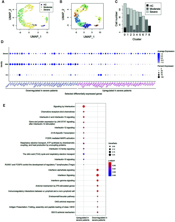
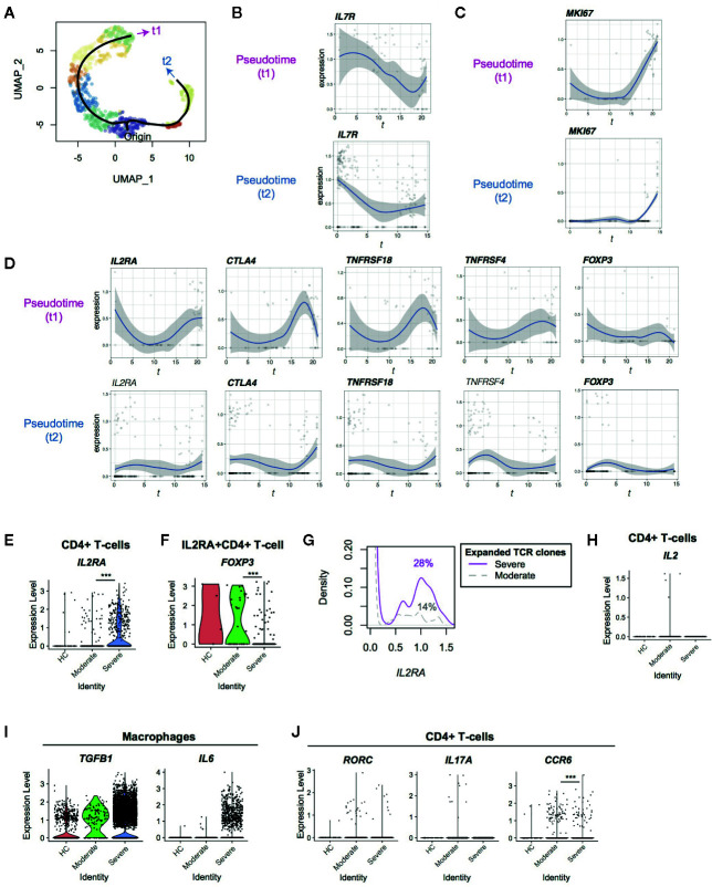
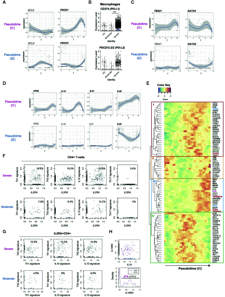
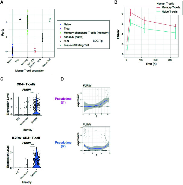
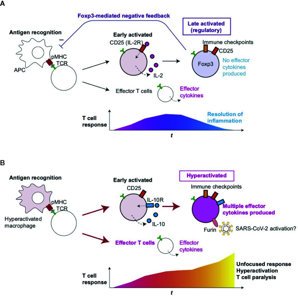
Severe COVID-19 patients show various immunological abnormalities including T-cell reduction and cytokine release syndrome, which can be fatal and is a major concern of the pandemic. However, it is poorly understood how T-cell dysregulation can contribute to the pathogenesis of severe COVID-19. Here we show single cell-level mechanisms for T-cell dysregulation in severe COVID-19, demonstrating new pathogenetic mechanisms of T-cell activation and differentiation underlying severe COVID-19. By in silico sorting CD4+ T-cells from a single cell RNA-seq dataset, we found that CD4+ T-cells were highly activated and showed unique differentiation pathways in the lung of severe COVID-19 patients. Notably, those T-cells in severe COVID-19 patients highly expressed immunoregulatory receptors and CD25, whilst repressing the expression of FOXP3. Furthermore, we show that CD25+ hyperactivated T-cells differentiate into multiple helper T-cell lineages, showing multifaceted effector T-cells with Th1 and Th2 characteristics. Lastly, we show that CD25-expressing hyperactivated T-cells produce the protease Furin, which facilitates the viral entry of SARS-CoV-2. Collectively, CD4+ T-cells from severe COVID-19 patients are hyperactivated and FOXP3-mediated negative feedback mechanisms are impaired in the lung, which may promote immunopathology. Therefore, our study proposes a new model of T-cell hyperactivation and paralysis that drives immunopathology in severe COVID-19.

Keywords: CD25; COVID-19; FOXP3; Furin; SARS-CoV-2; T-cells; regulatory T-cells (Tregs); single cell RNA-seq.


Figures

Figure 1
Figure 1
Single cell transcriptomes of CD4+ T-cells from COVID-19 patients. (A) UMAP analysis of in silico sorted CD4+ T-cells from COVID-19 patients. The colour code indicates the groups of patients: healthy control (HC), moderate and severe COVID-19 patients. (B) Clustering of single cells in the UMAP space, showing 9 Clusters (Clusters 0–8). (C) The proportion of single cells from each group in each cluster. (D) Dot plots showing the expression of selected differentially expressed genes between severe and moderate patients. (E) Pathway analysis of the differentially expressed genes.
Figure 2
Figure 2
Pseudotime analysis of CD4+ T-cells from Covid-19 patients for Treg-associated genes. (A) Two pseudotime trajectories were identified in the UMAP space. (B, C) The expression of (B) IL7R and (C) MKI67 in the pseudotime trajectories. (D) Gene expression dynamics of Treg-associated genes in the pseudotime trajectories. Genes with significant changes across pseudotime are highlighted by bold text. (E, F) The expression of IL2RA in (E) CD4+ T-cells and (F) IL2RA+CD4+ T-cells from the three groups. (G) IL2RA expression in CD4+T-cells with expanded TCR clones (n ≥ 2) in severe patients (magenta, solid line) and moderate patients (grey, broken line). Numbers indicate the percentage of IL2RA+ cells in each group. (H) The expression of IL2 in CD4+ T-cells from the three groups. (I) The expression of TGFB1 and IL6 in macrophages from the three groups. (J) The expression of the Th17 genes including RORC, IL17A, and CCR6 in CD4+ T-cells from the three groups. *** means p < .001.
Figure 3
Figure 3
Analysis of the expression dynamics of effector T-cell genes in CD4+ T-cells from COVID-19 patients. (A) The expression of BCL6 and PDCD1 in the pseudotime trajectories. (B) The expression of PD1 ligand genes, PD-L1 (CD274) and PD-L2 (PDCDLG2), in macrophages from the three groups. (C) The expression of Th1 transcription factor, TBX21, and Th2 transcription factor, GATA3, in the pseudotime trajectories. (D) Gene expression dynamics of IFNG, IL10, IL21 and IL32 in the pseudotime trajectories. (E) Heatmap analysis of selected genes in pseudotime 1 (t1). Here the rows are key genes that are differentially expressed across pseudotime 1, and the columns are single cells in the order of pseudotime 1. Th1, Th2, and Treg-associated genes are highlighted by cyan, green, and red. In addition, genes with significant changes across pseudotime are highlighted by bold text. (F, G) The expression of signature genes in (F) CD4+ T-cells and (G) IL2RA+CD4+ T-cells. (H) (upper panel) The expression of IL10 and IL10RA in CD4+ T-cells from COVID-19 patients. IL10+IL10RA+ double positive cells are highlighted by purple, while IL10+ single positive cells are shown by blue. (lower panel) Density plots of IL10RA expression in IL10+CD4+ T-cells and IL10-CD4+ T-cells.
Figure 4
Figure 4
FURIN expression in activated CD4+ T-cells in normal conditions and COVID-19 infection. (A) FURIN expression in CD4+ T-cell subpopulations from normal mice: naïve T-cells (naïve), Tregs, memory-phenotype T-cells (memory) from WT mice; non-draining lymph nodes (non-dLN, naïve), draining lymph nodes (dLN) of the pancreas, and pancreas-infiltrating effector T-cells (tissue-infiltrating Teff) of diabetes-prone BDC transgenic (Tg) mice. (B) Time course analysis of FURIN expression in human memory T-cells and naïve T-cells. (C) The expression of FURIN in CD4+ T-cells (upper) and IL2RA+CD4+ T-cells (lower) from the groups of patients and HC. (D) Gene expression dynamics of FURIN in the pseudotime trajectories 1 and 2.
Figure 5
Figure 5
Roles of T-cell hyperactivation in the lung of severe COVID-19 patients. (A) Peripheral Treg differentiation in normal conditions. Antigen-presenting cells (APC) present antigens as peptide-MHC complex (pMHC) to CD4+ T-cells, which triggers TCR signaling and subsequent activation and differentiation processes. Initially, early activated T-cells start to produce CD25 and IL-2, establishing a positive feedback loop for T-cell activation and proliferation. Some T-cells can differentiate into effector T-cells such as Th1 and Th2. Since IL-2 signaling enhances FOXP3 transcription, prolonged activation results in the expression of immune checkpoints such as CTLA-4 and FOXP3, which represses the transcription of effector cytokine genes. CD25+CTLA-4+FOXP3+ T-cells can consume and occupy immunological resources including IL-2 and CD28 signaling, and thereby mediate a negative feedback loop on the initial T-cell activation (27). This leads to the suppression of T-cell responses and the resolution of inflammation. (B) In severe COVID-19 patients, hyperactivated macrophages (13) may present antigens to CD4+ T-cells, which are activated and differentiate into CD25+ IL10R+ early activated T-cells which produce IL-10 rather than IL-2. FOXP3 transcription remains to be suppressed due to this and other unidentified mechanisms such as metabolism, while cytokines such as IL-10 further enhance the activation of CD25+ T-cells, resulting in the generation of CD25+ hyperactivated T-cells that express immune checkpoints, multiple effector T-cell cytokines, and Furin. The multifaceted Th differentiation may lead to unfocused T-cell responses and thereby paralyze the T-cell system. In addition, Furin can activate the S-protein of SARS-CoV-2 and thereby enhance viral entry into lung epithelial cells.

Similar articles

Cited by

References

    1. Okazaki T, Okazaki IM. Stimulatory and Inhibitory Co-signals in Autoimmunity. Adv Exp Med Biol (2019) 1189:213–32. 10.1007/978-981-32-9717-3_8 - DOI - PubMed
    1. Bending D, Ono M. From stability to dynamics: understanding molecular mechanisms of regulatory T cells through Foxp3 transcriptional dynamics. Clin Exp Immunol (2018) 197(1):14–23. 10.1111/cei.13194 - DOI - PMC - PubMed
    1. Hoffmann M, Kleine-Weber H, Schroeder S, Krüger N, Herrler T, Erichsen S, et al. SARS-CoV-2 Cell Entry Depends on ACE2 and TMPRSS2 and Is Blocked by a Clinically Proven Protease Inhibitor. Cell (2020) 181:271–280.e278. 10.1016/j.cell.2020.02.052 - DOI - PMC - PubMed
    1. Glowacka I, Bertram S, Muller M, Allen P, Soilleux E, Pfefferle S, et al. Evidence that TMPRSS2 Activates the Severe Acute Respiratory Syndrome Coronavirus Spike Protein for Membrane Fusion and Reduces Viral Control by the Humoral Immune Response. J Virol (2011) 85:4122–34. 10.1128/jvi.02232-10 - DOI - PMC - PubMed
    1. Sungnak W, Huang N, Bécavin C, Berg M, Queen R, Litvinukova M, et al. SARS-CoV-2 entry factors are highly expressed in nasal epithelial cells together with innate immune genes. Nat Med (2020) 26(5):681–7. 10.1038/s41591-020-0868-6 - DOI - PMC - PubMed

Publication types