. 2020 Oct 29:2020:8834360.
doi: 10.1155/2020/8834360. eCollection 2020.

Spinal Reflex Recovery after Dorsal Rhizotomy and Repair with Platelet-Rich Plasma (PRP) Gel Combined with Bioengineered Human Embryonic Stem Cells (hESCs)

Affiliations

Spinal Reflex Recovery after Dorsal Rhizotomy and Repair with Platelet-Rich Plasma (PRP) Gel Combined with Bioengineered Human Embryonic Stem Cells (hESCs)

Mateus Vidigal de Castro et al. Stem Cells Int. .

Abstract

Dorsal root rhizotomy (DRZ) is currently considered an untreatable injury, resulting in the loss of sensitive function and usually leading to neuropathic pain. In this context, we recently proposed a new surgical approach to treat DRZ that uses platelet-rich plasma (PRP) gel to restore the spinal reflex. Success was correlated with the reentry of primary afferents into the spinal cord. Here, aiming to enhance previous results, cell therapy with bioengineered human embryonic stem cells (hESCs) to overexpress fibroblast growth factor 2 (FGF2) was combined with PRP. For these experiments, adult female rats were submitted to a unilateral rhizotomy of the lumbar spinal dorsal roots, which was followed by root repair with PRP gel with or without bioengineered hESCs. One week after DRZ, the spinal cords were processed to evaluate changes in the glial response (GFAP and Iba-1) and excitatory synaptic circuits (VGLUT1) by immunofluorescence. Eight weeks postsurgery, the lumbar intumescences were processed for analysis of the repaired microenvironment by transmission electron microscopy. Spinal reflex recovery was evaluated by the electronic Von Frey method for eight weeks. The transcript levels for human FGF2 were over 37-fold higher in the induced hESCs than in the noninduced and the wildtype counterparts. Altogether, the results indicate that the combination of hESCs with PRP gel promoted substantial and prominent axonal regeneration processes after DRZ. Thus, the repair of dorsal roots, if done appropriately, may be considered an approach to regain sensory-motor function after dorsal root axotomy.


Conflict of interest statement

The authors declare that there is no conflict of interests regarding the publication of this paper.

Figures

Figure 1
Figure 1
(a–c) Cultivation of bioengineered human embryonic stem cell (hESC) clones over time. The formation of the cell monolayer (confluence) was observed on the fifth day (c). The cells were attached to a matrix (Matrigel) and expanded rapidly in their own medium, and they formed colonies and/or aggregates. Bright field. Scale bar = 10 μm. (d) Clones were induced to overexpress FGF2 at 48 hours after doxycycline administration. Practically all cells were fluorescent green (GFP+), which indicates that hESCs were activated by DOX and then overexpressed FGF2. Phase contrast. Scale bar = 25 μm. (e) A single bioengineered hESC was observed under electron microscopy, enabling visualization of the large nucleus, very evident nucleolus, and euchromatin, indicating high metabolic activity. Scale bar = 1 μm. (f–h) Relative expression of basic fibroblast growth factor (FGF2), brain-derived neurotrophic factor (BDNF), and glial cell line-derived neurotrophic factor (GDNF) mRNA in vitro. (f) The transcript levels for FGF2 were over 35-fold higher when modified hESCs were induced by doxycycline. Modified hESCs not treated with doxycycline and wildtype cells treated or not treated with doxycycline presented nearly undetectable expression of FGF2. (g) Compared with other groups, the transcript levels for BDNF were significantly higher when modified hESCs were induced by doxycycline. (h) The transcript levels for GDNF were not significantly different between groups. Mean ± SEM. ∗∗∗p < 0.001. hESCs: human embryonic stem cells; WT: wildtype; DOX: doxycycline.
Figure 2
Figure 2
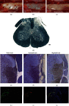
(a–c) Region of lumbar intumescence of an injury in a rat, seen through a surgical magnifying loupe. (a) Intact dorsal roots (L4, L5, and L6). (b) Transected dorsal roots (L4, L5, and L6). (c) PRP gel on the transected dorsal roots (L4, L5, and L6), immediately after its application. Immediately after applying the gel, it started to polymerize. From then on, it was no longer possible to reposition the roots that were transected on the spinal surface. Therefore, the repositioning of the roots after injury must be accurate before applying the gel. It is also possible to observe some hemorrhage after the lesion induction, where it was stopped with the gel. Scale bar = 5 mm. (d) Representative photomicrograph of transverse sections of the spinal cord stained with Sudan black 8 weeks after DRZ. The lesioned ipsilateral root was completely degenerated, as opposed to the contralateral root, which exhibits high integrity. Scale bar = 100 μm. (e–g) Photomicrograph of transverse sections of the hemispinal cord stained with toluidine blue, 8 weeks after the DRZ. (e) The unlesioned root showed a large number of axons. (f) The lesioned root was completely disrupted. (g) The repaired root showed a significant number of axons. Scale bar = 100 μm. (h–j) Bioengineered human embryonic stem cells (hESCs) found in the roots repaired with PRP, two weeks after injury: (h) hESCs overexpressing FGF2 (GFP+), in green; (i) hESCs marked with an antibody specific for human mitochondria (hMito), in red; (j) merge: GFP+ (green) + hMito (red), demonstrating that the engrafted cells are in fact the modified bioengineered hESCs. In blue, nuclear DNA labeling was performed using DAPI. Repaired root. ∗∗Spinal cord. The cells remained in the replanted root. Scale bar = 20 μm.
Figure 3
Figure 3
Electronic Von Frey measurements (mean values) obtained from the right hind paw (lesioned). The values are shown as the grams applied to trigger the “flinch” response. Statistical differences among groups are indicated with asterisks in addition to the graph. p < 0.05; ∗∗p < 0.01; ∗∗∗p < 0.001. Only the groups that underwent root repair with the PRP gel demonstrated the recovery of sensitivity to tactile stimulation. Further, the PRP gel + hESC group had the best performance in the whole experiment.
Figure 4
Figure 4
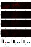
Representative immunofluorescence micrographs of VGLUT1 (glutamatergic synapses) immunolabeling on the ipsilateral side of the spinal cord after rhizotomy and repair, 1 week after lesion induction. A significant decrease in glutamatergic synaptic density was observed following DRZ. However, the “hESC” group showed great improvement in punctate labeling in comparison to the DRZ group. Scale bar = 50 μm.
Figure 5
Figure 5
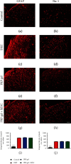
Representative immunofluorescence micrographs of GFAP (astrocytes) and Iba-1 (microglia) immunolabeling on the ipsilateral side of the spinal dorsal superficial laminae after lesion rhizotomy and repair. A significant increase in glial reactivity was observed after lesion induction. However, neither human PRP gel application nor hESC engrafting exacerbated glial reactivity, in comparison to the DRZ group. These findings directly reflected in the functional recovery from these groups. Scale bar = 50 μm.
Figure 6
Figure 6
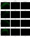
Representative immunofluorescence micrographs of CGRP immunolabeling on the ipsilateral side of the spinal cord after rhizotomy and repair, 1 week after lesion. Similar to VGLUT1 immunolabeling, a significant decrease in CGRP immunoreactivity was observed following DRZ. However, the “hESC” group showed a great improvement in CGRP-positive fibers in comparison to that of the DRZ group. Scale bar = 50 μm.
Figure 7
Figure 7
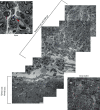
Representative transmission electronic micrographs of the repaired root and spinal cord dorsal horn (superficial region), 8 weeks after dorsal rhizotomy (DRZ) and repair with PRP gel. The extent of the repaired root can be observed with a significant number of myelinated axons (blue arrow), blood vessels (red arrow), and many Schwann cells (purple arrow). The image also highlights the subtle boundary between PNS/CNS clearly delimited by the dorsal root transition zone (DREZ), which contains central and peripheral nervous tissue. Moreover, proximal to the DREZ, myelin sheaths formed by oligodendrocytes can be observed, while further from the DREZ, the sheaths are formed by Schwann cells and wrapped the endoneurium. Their presence on the spinal surface of the root repaired in these morphological and structural conditions is one of the requirements for functional recovery.

Similar articles

Cited by

References

    1. Carlstedt T. New treatments for spinal nerve root avulsion injury. Frontiers in Neurology. 2016;7:p. 135. doi: 10.3389/fneur.2016.00135. - DOI - PMC - PubMed
    1. Scott S. H. The computational and neural basis of voluntary motor control and planning. Trends in Cognitive Sciences. 2012;16(11):541–549. doi: 10.1016/j.tics.2012.09.008. - DOI - PubMed
    1. Chew D. J., Murrell K., Carlstedt T., Shortland P. J. Segmental spinal root avulsion in the adult rat: a model to study avulsion injury pain. Journal of Neurotrauma. 2013;30(3):160–172. doi: 10.1089/neu.2012.2481. - DOI - PubMed
    1. Wu L., Wu J., Chang H. H., Havton L. A. Selective plasticity of primary afferent innervation to the dorsal horn and autonomic nuclei following lumbosacral ventral root avulsion and reimplantation in long term studies. Experimental Neurology. 2012;233(2):758–766. doi: 10.1016/j.expneurol.2011.11.034. - DOI - PubMed
    1. Ashley J., Budnik V. A tale of two inputs. Neuron. 2017;93(6):1245–1247. doi: 10.1016/j.neuron.2017.03.013. - DOI - PubMed