

Polygonally Meshed Dipole Model Simulation of the Electrical Field Produced by the Stomach and Intestines
- PMID: 33178331
- PMCID: PMC7607902
- DOI: 10.1155/2020/2971358
Polygonally Meshed Dipole Model Simulation of the Electrical Field Produced by the Stomach and Intestines
Abstract
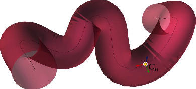
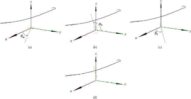
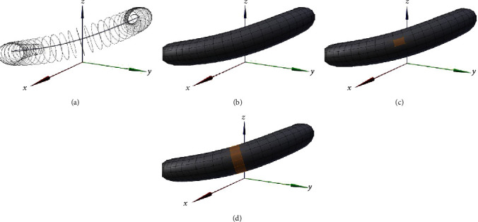
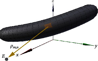
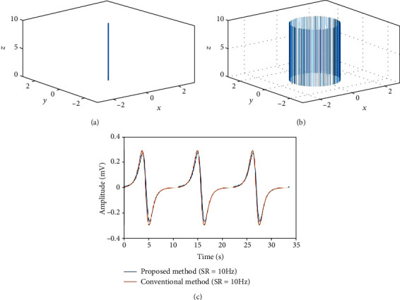
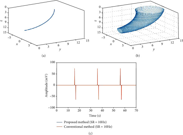
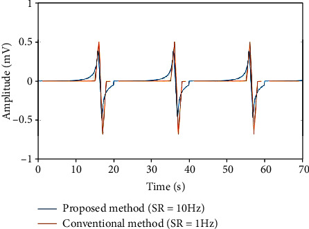
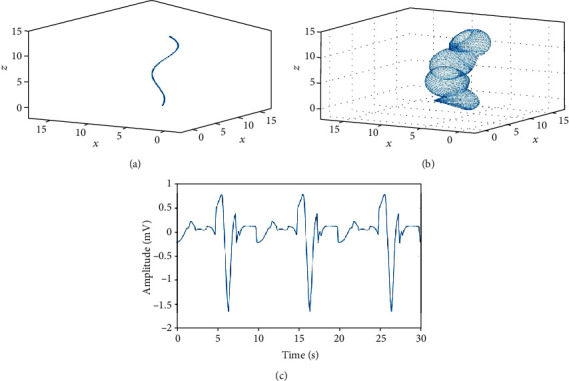
Cutaneous electrogastrography (EGG) is used in clinical and physiological fields to noninvasively measure the electrical activity of the stomach and intestines. Dipole models that mathematically express the electrical field characteristics generated by the stomach and intestines have been developed to investigate the relationship between the electrical control activity (ECA) (slow waves) shown in EGG and the internal gastric electrical activity. However, these models require a mathematical description of the movement of an annular band of dipoles, which limits the shape that can be modeled. In this study, we propose a novel polygonally meshed dipole model to conveniently reproduce ECA based on the movement of the annular band in complex shapes, such as the shape of the stomach and intestines, constructed in three-dimensional (3D) space. We show that the proposed model can reproduce ECA simulation results similar to those obtained using conventional models. Moreover, we show that the proposed model can reproduce the ECA produced by a complex geometrical shape, such as the shape of the intestines. The study results indicate that ECA simulations can be conducted based on structures that more closely resemble real organs than those used in conventional dipole models, with which, because of their intrinsic construction, it would be difficult to include realistic complex shapes, using the mathematical description of the movement of an annular band of dipoles. Our findings provide a powerful new approach for computer simulations based on the electric dipole model.
Copyright © 2020 Masaki Kawano and Takahiro Emoto.
Conflict of interest statement
The authors declare that there is no conflict of interest regarding the publication of this paper.
Figures
Similar articles
-
Conoidal dipole model of electrical field produced by the human stomach.Med Biol Eng Comput. 1995 Mar;33(2):179-84. doi: 10.1007/BF02523038. Med Biol Eng Comput. 1995. PMID: 7643657
-
A spatio-temporal dipole simulation of gastrointestinal magnetic fields.IEEE Trans Biomed Eng. 2003 Jul;50(7):836-47. doi: 10.1109/TBME.2003.813549. IEEE Trans Biomed Eng. 2003. PMID: 12848351
-
What is measured in electrogastrography?Dig Dis Sci. 1980 Mar;25(3):179-87. doi: 10.1007/BF01308136. Dig Dis Sci. 1980. PMID: 7371462
-
Anatomically realistic multiscale models of normal and abnormal gastrointestinal electrical activity.World J Gastroenterol. 2007 Mar 7;13(9):1378-83. doi: 10.3748/wjg.v13.i9.1378. World J Gastroenterol. 2007. PMID: 17457969 Free PMC article. Review.
-
Electrogastrography: measurement, analysis and prospective applications.Med Biol Eng Comput. 1991 Jul;29(4):339-50. doi: 10.1007/BF02441653. Med Biol Eng Comput. 1991. PMID: 1787748 Review.
References
-
- Alvarez W. C. The electrogastrogram and what it shows. JAMA: The Journal of the American Medical Association. 1922;78(15):1116–1119. doi: 10.1001/jama.1922.02640680020008. - DOI
-
- Cheng L. K., O’Grady G., Du P., Egbuji J. U., Windsor J. A., Pullan A. J. Detailed measurements of gastric electrical activity and their implications on inverse solutions. 2009 Annual International Conference of the IEEE Engineering in Medicine and Biology Society; September 2009; Minneapolis, MN, USA. pp. 1302–1305. - DOI - PMC - PubMed
MeSH terms
LinkOut - more resources
Full Text Sources
