

Fusion of whole and part features for the classification of histopathological image of breast tissue
- PMID: 33178434
- PMCID: PMC7642126
- DOI: 10.1007/s13755-020-00131-7
Fusion of whole and part features for the classification of histopathological image of breast tissue
Abstract
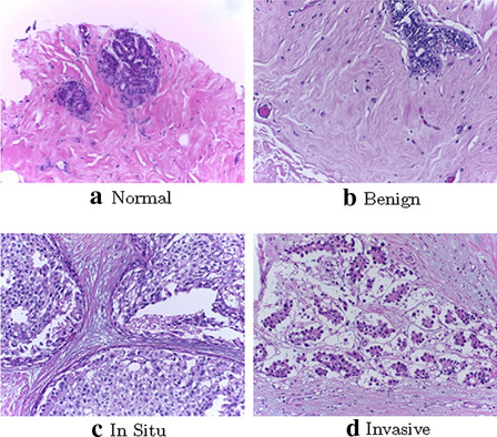
Purpose: Nowadays Computer-Aided Diagnosis (CAD) models, particularly those based on deep learning, have been widely used to analyze histopathological images in breast cancer diagnosis. However, due to the limited availability of such images, it is always tedious to train deep learning models that require a huge amount of training data. In this paper, we propose a new deep learning-based CAD framework that can work with less amount of training data.
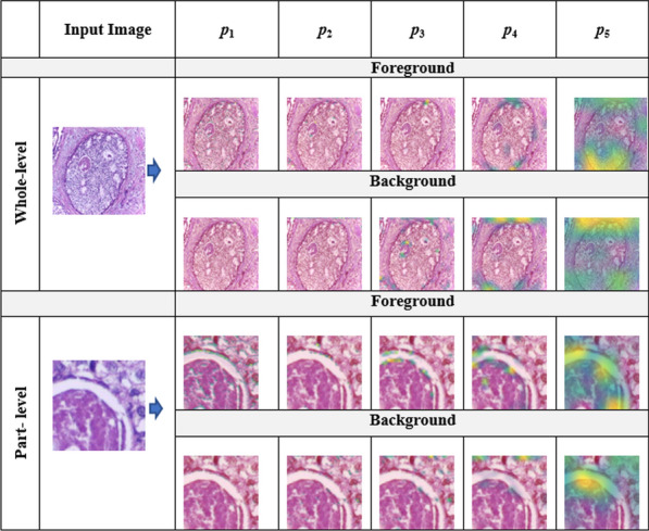
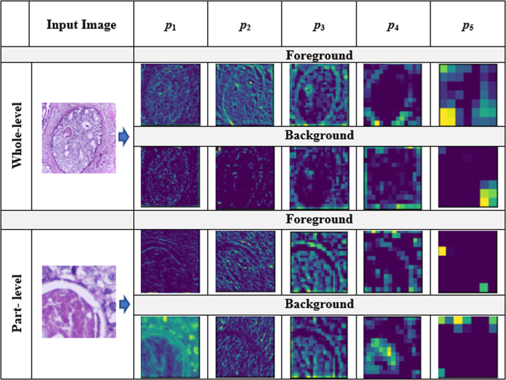
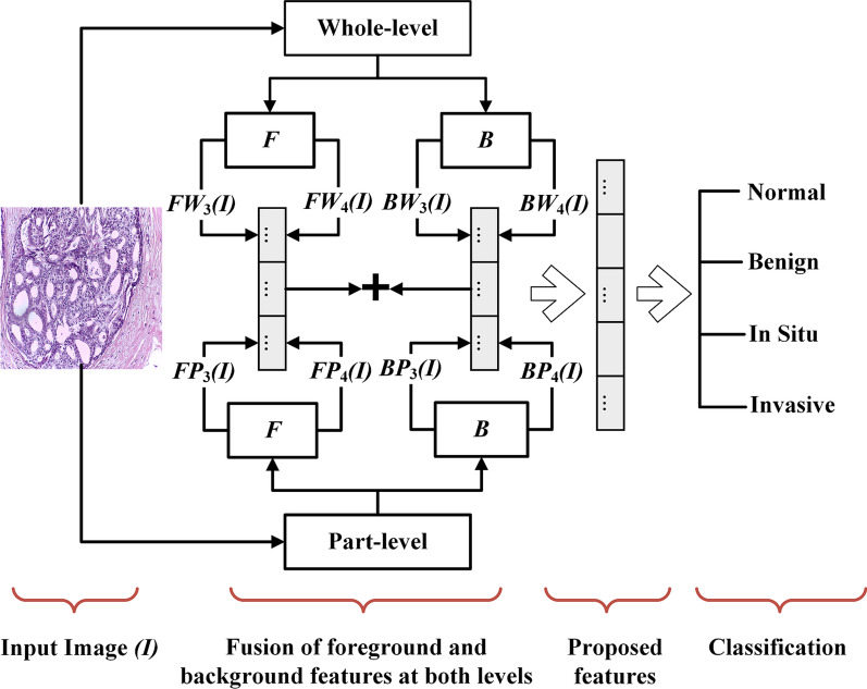
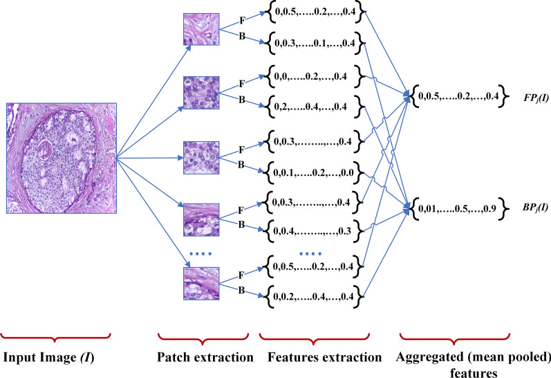
Methods: We use pre-trained models to extract image features that can then be used with any classifier. Our proposed features are extracted by the fusion of two different types of features (foreground and background) at two levels (whole-level and part-level). Foreground and background feature to capture information about different structures and their layout in microscopic images of breast tissues. Similarly, part-level and whole-level features capture are useful in detecting interesting regions scattered in high-resolution histopathological images at local and whole image levels. At each level, we use VGG16 models pre-trained on ImageNet and Places datasets to extract foreground and background features, respectively. All features are extracted from mid-level pooling layers of such models.
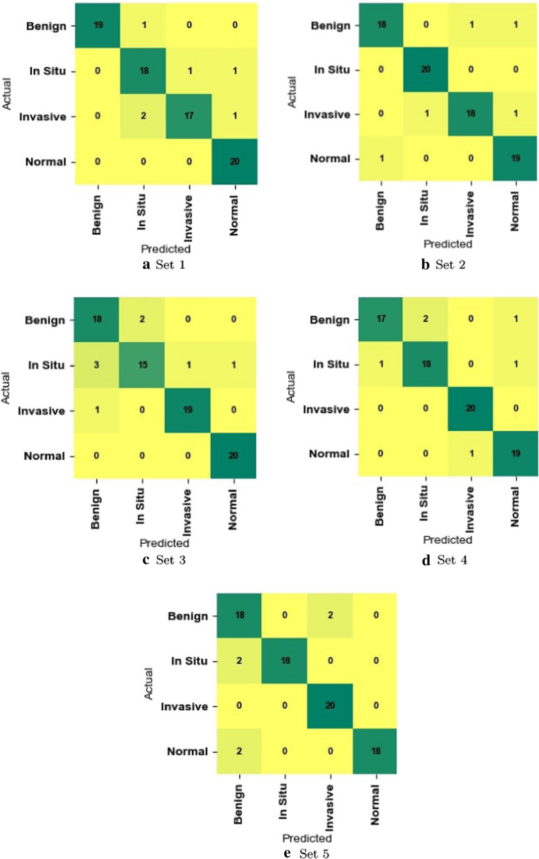
Results: We show that proposed fused features with a Support Vector Classifier (SVM) produce better classification accuracy than recent methods on BACH dataset and our approach is orders of magnitude faster than the best performing recent method (EMS-Net).
Conclusion: We believe that our method would be another alternative in the diagnosis of breast cancer because of performance and prediction time.
Keywords: Breast cancer; Computer-aided diagnosis; Deep learning; Histology; Histopathological images; Image classification.
© Springer Nature Switzerland AG 2020.
Conflict of interest statement
Conflict of interestWe would like to confirm that there are no known conflict of interests exist.
Figures
References
-
- Breiman L. Random forests. Mach Learn. 2001;45(1):5–32. doi: 10.1023/A:1010933404324. - DOI
-
- Campanella G, Silva VWK, Fuchs TJ. Terabyte-scale deep multiple instance learning for classification and localization in pathology. arXiv preprint. arXiv:180506983 (2018).
LinkOut - more resources
Full Text Sources
Miscellaneous
