

Tumor Cellular and Microenvironmental Cues Controlling Invadopodia Formation
- PMID: 33178698
- PMCID: PMC7593604
- DOI: 10.3389/fcell.2020.584181
Tumor Cellular and Microenvironmental Cues Controlling Invadopodia Formation
Abstract
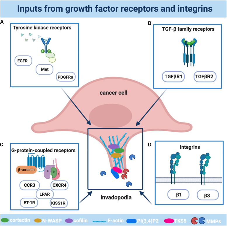
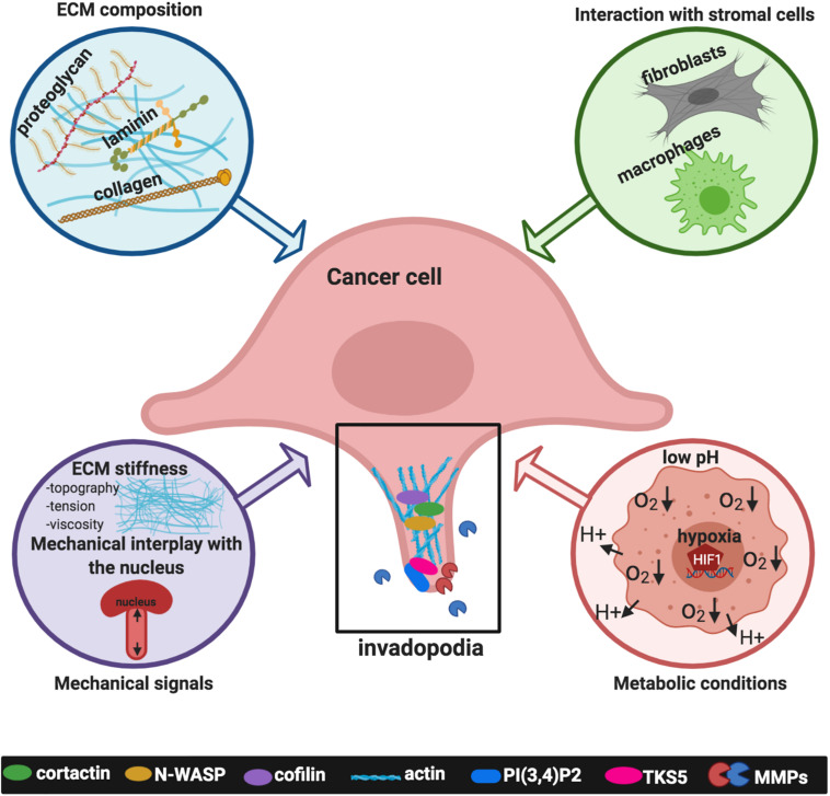
During the metastatic progression, invading cells might achieve degradation and subsequent invasion into the extracellular matrix (ECM) and the underlying vasculature using invadopodia, F-actin-based and force-supporting protrusive membrane structures, operating focalized proteolysis. Their formation is a dynamic process requiring the combined and synergistic activity of ECM-modifying proteins with cellular receptors, and the interplay with factors from the tumor microenvironment (TME). Significant advances have been made in understanding how invadopodia are assembled and how they progress in degradative protrusions, as well as their disassembly, and the cooperation between cellular signals and ECM conditions governing invadopodia formation and activity, holding promise to translation into the identification of molecular targets for therapeutic interventions. These findings have revealed the existence of biochemical and mechanical interactions not only between the actin cores of invadopodia and specific intracellular structures, including the cell nucleus, the microtubular network, and vesicular trafficking players, but also with elements of the TME, such as stromal cells, ECM components, mechanical forces, and metabolic conditions. These interactions reflect the complexity and intricate regulation of invadopodia and suggest that many aspects of their formation and function remain to be determined. In this review, we will provide a brief description of invadopodia and tackle the most recent findings on their regulation by cellular signaling as well as by inputs from the TME. The identification and interplay between these inputs will offer a deeper mechanistic understanding of cell invasion during the metastatic process and will help the development of more effective therapeutic strategies.
Keywords: cell invasion; cytoskeleton; extracellular matrix; invadopodia; metastasis; receptors; tumor microenvironment.
Copyright © 2020 Masi, Caprara, Bagnato and Rosanò.
Figures
Similar articles
-
Regulation of invadopodia by mechanical signaling.Exp Cell Res. 2016 Apr 10;343(1):89-95. doi: 10.1016/j.yexcr.2015.10.038. Epub 2015 Nov 4. Exp Cell Res. 2016. PMID: 26546985 Free PMC article. Review.
-
Quantitative measurement of invadopodia-mediated extracellular matrix proteolysis in single and multicellular contexts.J Vis Exp. 2012 Aug 27;(66):e4119. doi: 10.3791/4119. J Vis Exp. 2012. PMID: 22952016 Free PMC article.
-
Proteolytic and mechanical remodeling of the extracellular matrix by invadopodia in cancer.Phys Biol. 2022 Nov 21;20(1):10.1088/1478-3975/aca0d8. doi: 10.1088/1478-3975/aca0d8. Phys Biol. 2022. PMID: 36343366 Free PMC article. Review.
-
Dynamic interactions of cortactin and membrane type 1 matrix metalloproteinase at invadopodia: defining the stages of invadopodia formation and function.Cancer Res. 2006 Mar 15;66(6):3034-43. doi: 10.1158/0008-5472.CAN-05-2177. Cancer Res. 2006. PMID: 16540652
-
Regulation of invadopodia by the tumor microenvironment.Cell Adh Migr. 2014;8(3):226-35. doi: 10.4161/cam.28346. Cell Adh Migr. 2014. PMID: 24714597 Free PMC article. Review.
Cited by
-
Microfluidic Applications in Prostate Cancer Research.Micromachines (Basel). 2024 Sep 27;15(10):1195. doi: 10.3390/mi15101195. Micromachines (Basel). 2024. PMID: 39459070 Free PMC article. Review.
-
Invadopodia in cancer metastasis: dynamics, regulation, and targeted therapies.J Transl Med. 2025 May 16;23(1):548. doi: 10.1186/s12967-025-06526-y. J Transl Med. 2025. PMID: 40380267 Free PMC article. Review.
-
ADCYAP1 as a pan-solid cancer biomarker: predictor of immunotherapy efficacy in bladder cancer and prognostic potential across solid tumors.Discov Oncol. 2025 Apr 23;16(1):593. doi: 10.1007/s12672-025-02408-3. Discov Oncol. 2025. PMID: 40266460 Free PMC article.
-
Dual-specificity phosphatase 23 functions as a promising prognostic biomarker in non-small cell lung cancer.Genes Genomics. 2025 Mar;47(3):321-329. doi: 10.1007/s13258-024-01604-9. Epub 2024 Dec 18. Genes Genomics. 2025. PMID: 39693003
-
DCLK1-Mediated Regulation of Invadopodia Dynamics and Matrix Metalloproteinase Trafficking Drives Invasive Progression in Head and Neck Squamous Cell Carcinoma.bioRxiv [Preprint]. 2024 Apr 12:2024.04.06.588339. doi: 10.1101/2024.04.06.588339. bioRxiv. 2024. Update in: Mol Cancer. 2025 Feb 24;24(1):50. doi: 10.1186/s12943-025-02264-3. PMID: 38645056 Free PMC article. Updated. Preprint.
References
Publication types
LinkOut - more resources
Full Text Sources