Review
. 2020 Oct 16:8:597627.
doi: 10.3389/fcell.2020.597627. eCollection 2020.

Coreceptors and TCR Signaling - the Strong and the Weak of It

Affiliations
Review

Coreceptors and TCR Signaling - the Strong and the Weak of It

Alexander M Mørch et al. Front Cell Dev Biol. .

Abstract

The T-cell coreceptors CD4 and CD8 have well-characterized and essential roles in thymic development, but how they contribute to immune responses in the periphery is unclear. Coreceptors strengthen T-cell responses by many orders of magnitude - beyond a million-fold according to some estimates - but the mechanisms underlying these effects are still debated. T-cell receptor (TCR) triggering is initiated by the binding of the TCR to peptide-loaded major histocompatibility complex (pMHC) molecules on the surfaces of other cells. CD4 and CD8 are the only T-cell proteins that bind to the same pMHC ligand as the TCR, and can directly associate with the TCR-phosphorylating kinase Lck. At least three mechanisms have been proposed to explain how coreceptors so profoundly amplify TCR signaling: (1) the Lck recruitment model and (2) the pseudodimer model, both invoked to explain receptor triggering per se, and (3) two-step coreceptor recruitment to partially triggered TCRs leading to signal amplification. More recently it has been suggested that, in addition to initiating or augmenting TCR signaling, coreceptors effect antigen discrimination. But how can any of this be reconciled with TCR signaling occurring in the absence of CD4 or CD8, and with their interactions with pMHC being among the weakest specific protein-protein interactions ever described? Here, we review each theory of coreceptor function in light of the latest structural, biochemical, and functional data. We conclude that the oldest ideas are probably still the best, i.e., that their weak binding to MHC proteins and efficient association with Lck allow coreceptors to amplify weak incipient triggering of the TCR, without comprising TCR specificity.

Keywords: CD4; CD8; T-cell signaling; TCR triggering; antigen discrimination.


Figures

FIGURE 1
FIGURE 1
(A) T cells interact with functionalized lipid bilayers using multiple microvilli, forming either (B) a radially symmetric immunological synapse or (C) an asymmetrical, motile kinapse. These structures consist of organized SMAC domains which correspond to the underlying actin networks, indicated by color. Effector vesicles/particles are indicated by small membrane-bound circles. The kinapse is the primary behavior adopted by most human T cells stimulated by antigen with the exception of CD8+ memory T cells which are more likely to form stable synapses.
FIGURE 2
FIGURE 2
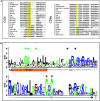
The coreceptor “zinc clasp” is highly conserved. (A) MUSCLE alignment of C-terminal CD4 and CD8α sequences with clasp cysteines highlighted in yellow and histidines in equivalent positions highlighted in blue. Adapted from Chida et al. (2011). (B) A sequence LOGO of the CD4 transmembrane (TM) helix and intracellular domain (ICD). Green triangles indicate glycines in the conserved GGXXG motif, blue triangles indicate S-palmitoylation sites, and red triangles indicate clasp cysteines. Sheep CD4 was excluded from this analysis due to the presence of large insertions in this regions bearing no homology to any other species.
FIGURE 3
FIGURE 3
Models for coreceptor function. (A) The Lck recruitment model: coreceptors recruit Lck to cognate pMHC-TCR complexes. (B) The pseudodimer model: coreceptors cross-link agonist-bound TCR to self-bound TCR. (C) The coreceptor recruitment model: ITAMs are incipiently phosphorylated by free Lck prior to recruitment of a coreceptor/Lck complex through SH2 domain-dependent interactions, e.g., between Lck and phosphorylated ITAMs. (D) The coreceptor scanning model: a cognate pMHC-TCR complex scans multiple “empty” coreceptors before encountering coreceptor-bound Lck. Gray arrows denote the passage of time. Only the zeta chain ITAMs are shown for simplicity. Protein models were generated from the crystal structures of TCR/CD3 (PDB ID: 6JXR), CD4 (PDB ID: 1WIQ), HLA-DR1 (PDB ID: 4I5B), and the ternary TCR-pMHC-CD4 complex (PDB ID: 3T0E).

Similar articles

Cited by

References

    1. Arcaro A., Grégoire C., Bakker T. R., Baldi L., Jordan M., Goffin L., et al. (2001). CD8β endows CD8 with efficient coreceptor function by coupling T cell receptor/CD3 to raft-associated CD8/p56lck complexes. J. Exp. Med. 194 1485–1495. 10.1084/jem.194.10.1485 - DOI - PMC - PubMed
    1. Arcaro A., Grégoire C., Boucheron N., Stotz S., Palmer E., Malissen B., et al. (2000). Essential role of CD8 palmitoylation in CD8 coreceptor function. J. Immunol. 165 2068–2076. 10.4049/jimmunol.165.4.2068 - DOI - PubMed
    1. Artyomov M. N., Lis M., Devadas S., Davis M. M., Chakraborty A. K. (2010). CD4 and CD8 binding to MHC molecules primarily acts to enhance Lck delivery. Proc. Natl. Acad. Sci. U.S.A. 107 16916–16921. 10.1073/pnas.1010568107 - DOI - PMC - PubMed
    1. Bach F. H., Bach M. L., Sondel P. M. (1976). Differential function of major histocompatibility complex antigens in T-lymphocyte activation. Nature 259 273–281. 10.1038/259273a0 - DOI - PubMed
    1. Balint Š, Müller S., Fischer R., Kessler B. M., Harkiolaki M., Valitutti S., et al. (2020). Supramolecular attack particles are autonomous killing entities released from cytotoxic T cells. Science 368:eaay9207. 10.1126/science.aay9207 - DOI - PMC - PubMed