Review
. 2019 Mar 8;1(1):20180049.
doi: 10.1259/bjro.20180049. eCollection 2019.

Diffusion-weighted imaging of the breast: current status as an imaging biomarker and future role

Affiliations
Review

Diffusion-weighted imaging of the breast: current status as an imaging biomarker and future role

Julia Camps-Herrero. BJR Open. .

Abstract

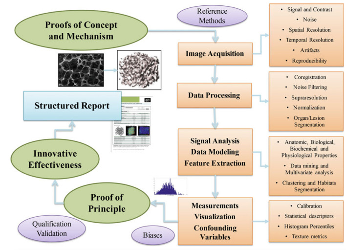
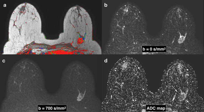
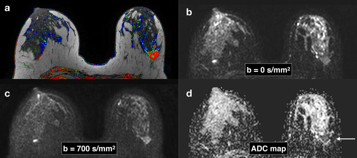
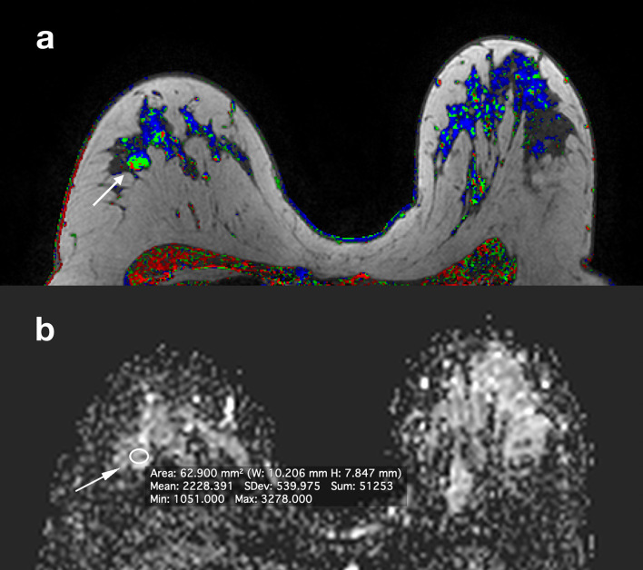
Diffusion-weighted imaging (DWI) of the breast is a MRI sequence that shows several advantages when compared to the dynamic contrast-enhanced sequence: it does not need intravenous contrast, it is relatively quick and easy to implement (artifacts notwithstanding). In this review, the current applications of DWI for lesion characterization and prognosis as well as for response evaluation are analyzed from the point of view of the necessary steps to become a useful surrogate of underlying biological processes (tissue architecture and cellularity): from the proof of concept, to the proof of mechanism, the proof of principle and finally the proof of effectiveness. Future applications of DWI in screening, DWI modeling and radiomics are also discussed.


Figures

Figure 1.
Figure 1.
The stepwise development of imaging biomarkers. From Introduction to the Stepwise Development of Imaging Biomarkers (Chapter 2) in Imaging Biomarkers. Martí-Bonmatí L, Alberich-Bayarri A, editors. Springer; 2016. With permission of the author.
Figure 2.
Figure 2.
Breast images obtained with DWI in a patient with a Triple Negative subtype breast cancer in the left breast. Corresponding slices from DCE postcontrast image (a), DWI at b = 0 s/mm2 (b), DWI at b = 700 s/mm2. (c), ADC map (d). Invasive tumors show reduced diffusivity on DW imaging, appearing hyperintense on b = 700 s/mm2 image (c) and hypointense on the ADC map (arrows) (d). ADC, apparent diffusion coefficient; DCE, dynamiccontrast-enhanced; DWI, diffusion-weighted imaging.
Figure 3.
Figure 3.
Breast images obtained with DWI in a patient with a HER-2 subtype breast cancer in the left breast. Corresponding slices from DCE post-contrast image (a), DWI at b = 0 s/mm2 (b), DWI at b = 700 s/mm2 . (c), ADC map (d). Invasive tumors (arrows) show reduced diffusivity on DW imaging, appearing hyperintense on b = 700 s/mm2 image (c) and hypointense on the ADC map (arrow) (d). ADC, apparent diffusion coefficient; DCE, dynamic contrast-enhanced; DWI, diffusion-weighted imaging.
Figure 4.
Figure 4.
Same patient in Figure 2. Corresponding slices from DCE postcontrast image (a), ADC map (b). A BI-RADS four lesion (arrow) was identified in the contralateral breast and DWI characterized it as benign (ADC = 2,22×10⁻³ mm2/s). It was decided not to biopsy. 2 years later, the lesion was stable and 3 years later the patient underwent contralateral prophylactic mastectomy which did not show any malignant lesions. ADC, apparent diffusion coefficient; DCE, dynamic contrast-enhanced; DWI, diffusion-weighted imaging.
Figure 5.
Figure 5.
Same patient in Figure 2. Corresponding slices from DCE postcontrast image (a), DCE subtraction image (b) ADC map (c). Two BI-RADS four lesions (arrows) were identified in a different quadrant from the index tumor. ADC characterized them as malignant (ADC = 1,28×10⁻³ mm2/s). An MR-guided vacuum-assisted biopsy was performed in the largest of the two lesions and an additional multicentric invasive ductal cancer was confirmed. ADC, apparent diffusion coefficient; DCE, dynamiccontrast-enhanced; DWI, diffusion-weighted imaging.
Figure 6.
Figure 6.
Breast images obtained in a patient with a Triple Negative subtype breast cancer in the left breast (same patient from Figure 1): baseline pre-neoadjuvant chemotherapy, early after two cycles and pre-surgery after eight cycles. Slices from baseline, early (after two cycles) and pre-surgical (after eight cycles) DTI in the top row. Corresponding slices (bottom row) from baseline post-contrast image, T 2 weighted image after two cycles and post-contrast image after eight cycles, pre-surgery. Note that in the DTI images after two cycles, there is a functional qualitative partial major almost complete response whilst in the corresponding T 2 weighted image, the morphologic response is minor or minimal. When comparing the DTI images after eight cycles with the corresponding DCE image, there is a complete response in the DTI images (arrow, top row) and a residual enhancement in the DCE images (arrow, bottom row) interpreted as partial major response. The final pathology confirmed a complete response that was already predicted by the early DTI exam after two cycles. DCE, dynamic contrast-enhanced; DTI, diffusion tensor imaging.

References

    1. European Society of Radiology (ESR) ESR statement on the stepwise development of imaging biomarkers. Insights Imaging 2013; 4: 147–52. doi: 10.1007/s13244-013-0220-5 - DOI - PMC - PubMed
    1. Martí-Bonmatí L. Introduction to the stepwise development of imaging biomarkers : Imaging Biomarkers. 30: The British Institute of Radiology.; 2016. 9–27.
    1. Paran Y, Bendel P, Margalit R, Degani H. Water diffusion in the different microenvironments of breast cancer. NMR Biomed 2004; 17: 170–80 2004. doi: 10.1002/nbm.882 - DOI - PubMed
    1. White NS, McDonald C, McDonald CR, Farid N, Kuperman J, Karow D, Schenker-Ahmed NM, et al. . Diffusion-weighted imaging in cancer: physical foundations and applications of restriction spectrum imaging. Cancer Res 2014; 74: 4638–52. doi: 10.1158/0008-5472.CAN-13-3534 - DOI - PMC - PubMed
    1. Le Bihan D, Breton E, Lallemand D, Grenier P, Cabanis E, Laval-Jeantet M. MR imaging of intravoxel incoherent motions: application to diffusion and perfusion in neurologic disorders. Radiology 1986; 161: 401–7. doi: 10.1148/radiology.161.2.3763909 - DOI - PubMed