Review
. 2019 Jun 24;1(1):20190001.
doi: 10.1259/bjro.20190001. eCollection 2019.

Imaging biomarkers in upper gastrointestinal cancers

Affiliations
Review

Imaging biomarkers in upper gastrointestinal cancers

Michela Gabelloni et al. BJR Open. .

Abstract

In parallel with the increasingly widespread availability of high performance imaging platforms and recent progresses in pathobiological characterisation and treatment of gastrointestinal malignancies, imaging biomarkers have become a major research topic due to their potential to provide additional quantitative information to conventional imaging modalities that can improve accuracy at staging and follow-up, predict outcome, and guide treatment planning in an individualised manner. The aim of this review is to briefly examine the status of current knowledge about imaging biomarkers in the field of upper gastrointestinal cancers, highlighting their potential applications and future perspectives in patient management from diagnosis onwards.


Figures

Figure 1.
Figure 1.
An 83-year-old male with locally advanced, partially necrotic gingival SCC as displayed on standard morphological CT image (a). Quantitative CT perfusion analysis revealed increased blood flow (BF) (b), blood volume (BV) (c), permeability surface (PS) product (d), and reduced mean transit time (MTT) (e) values in the viable portion of the tumour compared with contralateral normal tissue (Ref) taken as reference. Note lack of perfusion inside the necrotic portion of the tumour as visually depicted on colour-coded perfusion maps.
Figure 2.
Figure 2.
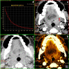
A 51-year-old female with small keratinising SCC of the left retromolar trigone (arrow on DECT monochromatic 55keV image, (a). Material decomposition iodine/water image (b) improves lesion conspicuity by boosting iodine signal and reveals high lesion vascularisation (measured mean iodine concentration of 32 × 100 µg ml−1), whereas virtual precontrast image (c) shows no abnormal density or beam hardening artefacts at the lesion site. Avid lesion enhancement was confirmed by spectral curve analysis (d). All images had been obtained at the same anatomic level from a single contrast-enhanced DECT acquisition.
Figure 3.
Figure 3.
A 49-year-old male with T3N2 advanced gastric cancer without peritoneal seeding showed entropy of 7.05 within the region of interest (a). A 59-year-old female with T3N2 advanced gastric cancer with occult seeding (b) showed entropy of 7.70, higher than the cut-off value (>7.141) obtained from receiver operating characteristic (ROC) curve analysis (c). Reproduced and adapted from.

References

    1. Califf RM. Biomarker definitions and their applications. Exp Biol Med 2018; 243: 213–21. doi: 10.1177/1535370217750088 - DOI - PMC - PubMed
    1. FDA-NIH Biomarker Working Group Best (biomarkers, endpoints. and other Tools) Resource 2018;Available from. - PubMed
    1. Deigner H. P, Kohl M. Precision medicine: tools and quantitative approaches. London, UK: The British Institute of Radiology.; 2018. .
    1. European Society of Radiology (ESR). white paper on imaging biomarkers. Insights Imaging 2010; 1: 42–5. - PMC - PubMed
    1. Lambin P, Rios-Velazquez E, Leijenaar R, Carvalho S, van Stiphout RGPM, Granton P, et al. . Radiomics: extracting more information from medical images using advanced feature analysis. Eur J Cancer 2012; 48: 441–6. doi: 10.1016/j.ejca.2011.11.036 - DOI - PMC - PubMed