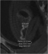
Biometric analysis of the foetal meconium pattern using T1 weighted 2D gradient echo MRI
- PMID: 33178986
- PMCID: PMC7594886
- DOI: 10.1259/bjro.20200032
Biometric analysis of the foetal meconium pattern using T1 weighted 2D gradient echo MRI
Abstract
Objectives: Foetal MRI is used to assess abnormalities after ultrasonography. Bowel anomalies are a significant cause of neonatal morbidity, however there are little data concerning its normal appearance on antenatal MRI. This study aims to investigate the pattern of meconium accumulation throughout gestation using its hyperintense appearance on T 1 weighted scans and add to the current published data.
Methods: This was a retrospective cohort study in a tertiary referral clinical MRI centre. Foetal body MRI scans of varying gestational ages were obtained dating between October 2011 and March 2018. The bowel was visualised on T 1 weighted images. The length of the meconium and the width of the meconium at the rectum, sigmoid colon, splenic flexure and hepatic flexure was measured. Presence or absence of meconium in the small bowel was noted. Inter- and intrarater reliability was assessed.
Results: 181 foetal body scans were reviewed. 52 were excluded and 129 analysed. Visualisation of the meconium in the large bowel became increasingly proximal with later gestations, and small bowel visualisation was greater at earlier gestations. There was statistically significant strong (r = 0.6-0.8) or very strong (r = 0.8-1.0) positive correlation of length and width with increasing gestation. Interrater reliability was moderate to excellent (r = 0.4-1.0).
Conclusion: This study provides new information regarding the pattern of meconium accumulation throughout gestation. With care, the results can be used in clinical practice to aid diagnosis of bowel pathology.
Advances in knowledge: The findings of this study provide further information concerning the normal accumulation of foetal meconium on MR imaging, an area where current research is limited.
© 2020 The Authors. Published by the British Institute of Radiology.
Conflict of interest statement
Competing interests: The authors declare no conflicts of interest. Competing interests: The authors declare no conflicts of interest.
Figures


Similar articles
-
Visualisation of fetal meconium on post-mortem magnetic resonance imaging scans: a retrospective observational study.Acta Radiol Open. 2020 Nov 19;9(11):2058460120970541. doi: 10.1177/2058460120970541. eCollection 2020 Nov. Acta Radiol Open. 2020. PMID: 33282338 Free PMC article.
-
Fetal MRI of urine and meconium by gestational age for the diagnosis of genitourinary and gastrointestinal abnormalities.AJR Am J Roentgenol. 2005 Jun;184(6):1891-7. doi: 10.2214/ajr.184.6.01841891. AJR Am J Roentgenol. 2005. PMID: 15908548
-
MRI of the fetal gastrointestinal tract.Pediatr Radiol. 2002 Jun;32(6):395-404. doi: 10.1007/s00247-001-0607-1. Epub 2002 Feb 16. Pediatr Radiol. 2002. PMID: 12029338
-
Fetal MR Imaging of Gastrointestinal Abnormalities.Radiographics. 2016 May-Jun;36(3):904-17. doi: 10.1148/rg.2016150109. Radiographics. 2016. PMID: 27163598 Review.
-
Fetal bowel anomalies--US and MR assessment.Pediatr Radiol. 2012 Jan;42 Suppl 1:S101-6. doi: 10.1007/s00247-011-2174-4. Epub 2012 Mar 6. Pediatr Radiol. 2012. PMID: 22395722 Review.
Cited by
-
Is that bowel normal? Nomograms for fetal colon and rectum measurements by MRI from 20-36 weeks' gestation.Pediatr Radiol. 2025 May;55(5):987-998. doi: 10.1007/s00247-025-06192-8. Epub 2025 Feb 20. Pediatr Radiol. 2025. PMID: 39976709
-
Standardised and structured reporting in fetal magnetic resonance imaging: recommendations from the Fetal Task Force of the European Society of Paediatric Radiology.Pediatr Radiol. 2024 Sep;54(10):1566-1578. doi: 10.1007/s00247-024-06010-7. Epub 2024 Aug 1. Pediatr Radiol. 2024. PMID: 39085531 Review.
References
-
- Reddy UM, Filly RA, Copel JA, .Pregnancy and Perinatology Branch, Eunice Kennedy Shriver National Institute of Child Health and Human Development, Department of Health and Human Services, NIH . Prenatal imaging: ultrasonography and magnetic resonance imaging. Obstet Gynecol 2008; 112: 145–57. doi: 10.1097/01.AOG.0000318871.95090.d9 - DOI - PMC - PubMed
LinkOut - more resources
Full Text Sources

