

Resective surgery prevents progressive cortical thinning in temporal lobe epilepsy
- PMID: 33179036
- PMCID: PMC7719024
- DOI: 10.1093/brain/awaa284
Resective surgery prevents progressive cortical thinning in temporal lobe epilepsy
Abstract
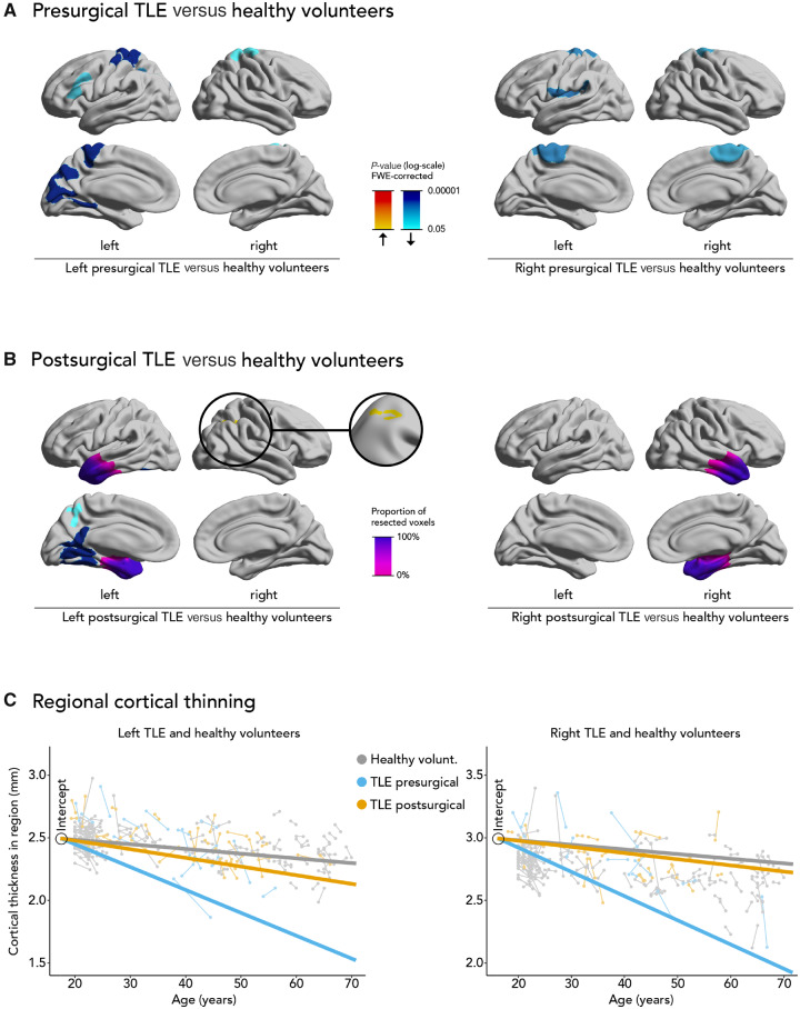
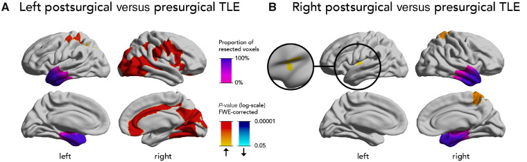
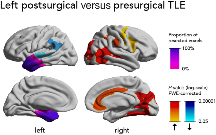
Focal epilepsy in adults is associated with progressive atrophy of the cortex at a rate more than double that of normal ageing. We aimed to determine whether successful epilepsy surgery interrupts progressive cortical thinning. In this longitudinal case-control neuroimaging study, we included subjects with unilateral temporal lobe epilepsy (TLE) before (n = 29) or after (n = 56) anterior temporal lobe resection and healthy volunteers (n = 124) comparable regarding age and sex. We measured cortical thickness on paired structural MRI scans in all participants and compared progressive thinning between groups using linear mixed effects models. Compared to ageing-related cortical thinning in healthy subjects, we found progressive cortical atrophy on vertex-wise analysis in TLE before surgery that was bilateral and localized beyond the ipsilateral temporal lobe. In these regions, we observed accelerated annualized thinning in left (left TLE 0.0192 ± 0.0014 versus healthy volunteers 0.0032 ± 0.0013 mm/year, P < 0.0001) and right (right TLE 0.0198 ± 0.0016 versus healthy volunteers 0.0037 ± 0.0016 mm/year, P < 0.0001) presurgical TLE cases. Cortical thinning in these areas was reduced after surgical resection of the left (0.0074 ± 0.0016 mm/year, P = 0.0006) or right (0.0052 ± 0.0020 mm/year, P = 0.0006) anterior temporal lobe. Directly comparing the post- versus presurgical TLE groups on vertex-wise analysis, the areas of postoperatively reduced thinning were in both hemispheres, particularly, but not exclusively, in regions that were affected preoperatively. Participants who remained completely seizure-free after surgery had no more progressive thinning than that observed during normal ageing. Those with postoperative seizures had small areas of continued accelerated thinning after surgery. Thus, successful epilepsy surgery prevents progressive cortical atrophy that is observed in TLE and may be potentially neuroprotective. This effect was more pronounced in those who remained seizure-free after temporal lobe resection, normalizing the rate of atrophy to that of normal ageing. These results provide evidence of epilepsy surgery preventing further cerebral damage and provide incentives for offering early surgery in refractory TLE.
Keywords: MRI; epilepsy; neurodegeneration; seizures; surgery.
© The Author(s) (2020). Published by Oxford University Press on behalf of the Guarantors of Brain. All rights reserved. For permissions, please email: journals.permissions@oup.com.
Figures

Comment in
-
Is bad brain worse than no brain? Salvaging the cerebral cortex in epilepsy.Brain. 2020 Dec 5;143(11):3172-3175. doi: 10.1093/brain/awaa330. Brain. 2020. PMID: 33278819 No abstract available.
-
Flattening the Curve: Slowing Age-Accelerated Brain Atrophy With Epilepsy Surgery.Epilepsy Curr. 2021 Mar 16;21(3):159-161. doi: 10.1177/15357597211001066. eCollection 2021 May-Jun. Epilepsy Curr. 2021. PMID: 34867092 Free PMC article. No abstract available.
References
-
- Alvim MKM, Coan AC, Campos BM, Yasuda CL, Oliveira MC, Morita ME, et al. Progression of gray matter atrophy in seizure-free patients with temporal lobe epilepsy. Epilepsia 2016; 57: 621–9. - PubMed
-
- Blümcke I, Spreafico R, Haaker G, Coras R, Kobow K, Bien CG, et al. Histopathological findings in brain tissue obtained during epilepsy surgery. N Engl J Med 2017; 377: 1648–56. - PubMed
