. 2021 Jan;23(1):22.
doi: 10.3892/mmr.2020.11660. Epub 2020 Nov 12.

Involvement of Klotho, TNF‑α and ADAMs in radiation‑induced senescence of renal epithelial cells

Affiliations

Involvement of Klotho, TNF‑α and ADAMs in radiation‑induced senescence of renal epithelial cells

Da Yeon Kim et al. Mol Med Rep. 2021 Jan.

Abstract

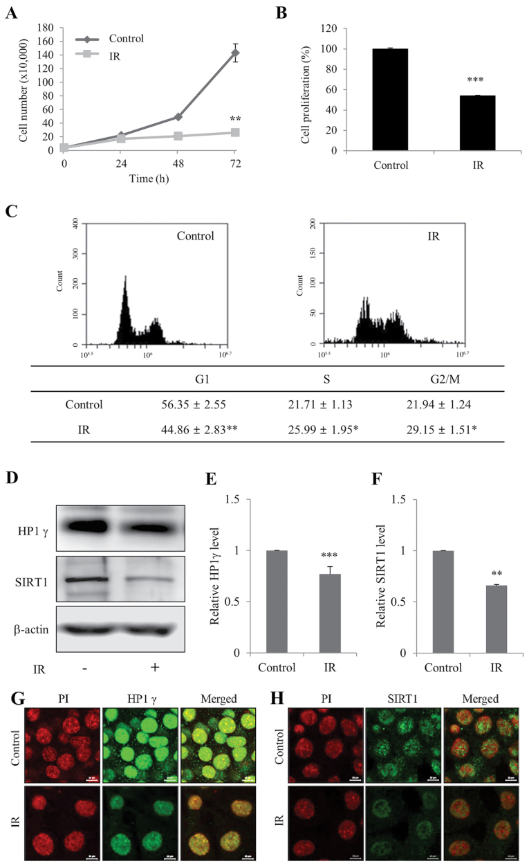
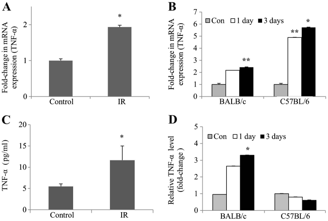
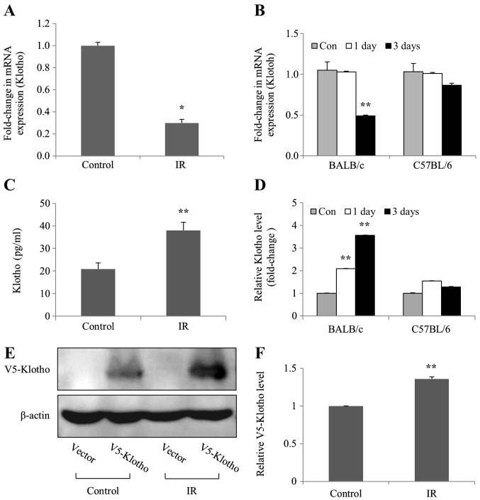
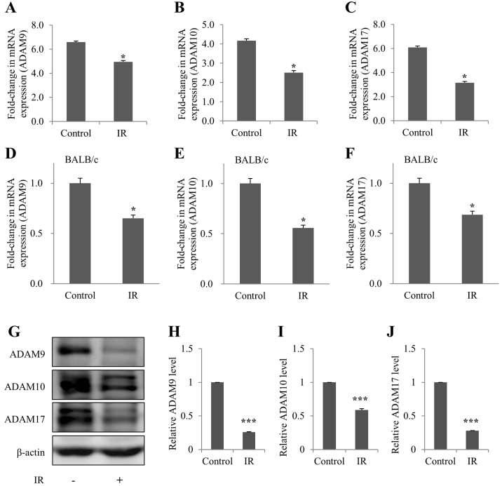
While radiation nephropathy is a major problem associated with radiotherapy, the exact mechanisms underlying its pathogenesis and the mediators involved in kidney deterioration remain to be elucidated. In view of the finding that senescence is typically increased post‑irradiation, the present study examined whether ionizing radiation may cause kidney injury by enhancing premature senescence. The present study explored the relevance of the aging suppressor, Klotho, which has anti‑aging activity and is highly expressed in murine renal cells/kidney tissues, under irradiation conditions. Firstly, the effects of radiation on mouse inner medullary collecting duct‑3 (mIMCD‑3) cells and kidney tissues of mice were assessed. Subsequently, the mRNA expression levels of Klotho, TNF‑α and ADAM metallopeptidase domain (ADAM)9/10/17 were analyzed by reverse transcription‑quantitative PCR following exposure to radiation. In addition, the levels of these proteins were measured by western blotting or ELISA. The results revealed that irradiation of mIMCD‑3 cells clearly triggered cellular senescence. Notably, Klotho gene expression was considerably decreased in radiation‑exposed mIMCD‑3 cells and in the kidney tissues of irradiated BALB/c mice, and the corresponding translated protein was consistently expressed following radiation exposure. Moreover, expression of TNF‑α, a negative regulator of Klotho, was significantly increased, whereas ADAM9/10/17, an ectodomain shedding enzyme of Klotho, was decreased in irradiated mIMCD‑3 cells and in the kidney tissues of BALB/c mice. Collectively, these data suggested that TNF‑α‑mediated inhibition of Klotho expression and blockage of soluble Klotho formation via decreased ADAM expression following irradiation may contribute to the development of renal dysfunction through acceleration of radiation‑induced cellular senescence.

Keywords: radiation; senescence; renal epithelial cells; Klotho; TNF‑α; ADAM metallopeptidase domain.


Figures

Figure 1.
Figure 1.
Radiation induces senescence of mIMCD-3 cells. (A) Control and IR-treated viable mIMCD-3 cells were visualized using Trypan blue exclusion and counted. (B) Control and IR-treated cell proliferation wase determined using the WST assay. Data are presented as a percentage of control cell growth. (C) Control and IR-treated mIMCD-3 cells were stained with PI and subjected to flow cytometry. (D) Protein expression levels of HP1γ and SIRT1 were determined via western blotting, with β-actin as the loading control. Semi-quantification of (E) HP1γ and (F) SIRT1 protein expression levels in cell lysates relative to the control group. Immunofluorescence analysis of (G) HP1γ and (H) SIRT1 using a fluorescein isothiocyanate-conjugated secondary antibody and PI nuclear staining followed by confocal microscopy (scale bar, 10 µm). mIMCD-3 cells were treated with or without 6 Gy radiation and cultured for 72 h. Data are expressed as the mean ± SD (n=4), *P<0.05, **P<0.01, ***P<0.001 vs. Control. mIMCD-3, mouse inner medullary collecting duct-3; IR, ionizing radiation; HP1γ, heterochromatin protein 1γ; SIRT1, sirtuin 1; PI, propidium iodide.
Figure 2.
Figure 2.
Upregulation of TNF-α mRNA expression and protein levels in irradiated mIMCD-3 cells, and renal tissues of BALB/c and C57BL6 mice. (A) mIMCD-3 cells, and (B) BALB/c and C57BL/6 mice were treated with or without 6 Gy radiation. After 24 and/or 72 h, reverse transcription-quantitative PCR was applied to assess TNF-α mRNA expression using GAPDH as the loading or internal control. ELISA was conducted to measure (C) protein levels of TNF-α (pg/ml) in mIMCD-3 cells, and (D) fold-change values of TNF-α levels in BALB/c and C57BL/6 mice relative to the control group. Data are presented as the mean ± SD (n=4). *P<0.05, **P<0.01 vs. Control. mIMCD-3, mouse inner medullary collecting duct-3; IR, ionizing radiation.
Figure 3.
Figure 3.
mRNA expression levels of Klotho are inhibited in mIMCD-3 cells, and renal tissues of BALB/c and C57BL6 mice after radiation exposure. (A) mIMCD-3 cells, and (B) BALB/c and C57BL/6 mice were treated with or without 6 Gy radiation. After 24 and/or 72 h, reverse transcription-quantitative PCR was applied to assess the mRNA expression levels of Klotho using GAPDH as the loading or internal control. ELISA was conducted to measure (C) Klotho protein levels (pg/ml) in mIMCD-3 cells, and (D) fold-change values of Klotho levels in BALB/c and C57BL/6 mice relative to the control group. (E) Following transient transfection with V5-tagged Klotho expression plasmid, mIMCD-3 cells were exposed to 6 Gy radiation and protein expression levels of Anti-V5 in cell lysates were determined via western blotting using Klotho-specific antibodies 24 h after irradiation. Variations in protein loading among samples were controlled by monitoring the β-actin level. (F) Semi-quantification of V5-tagged Klotho protein expression levels in IR-irradiated cell lysates relative to V5-tagged Klotho protein expression levels in control group. Data are presented as the mean ± SD (n=4). *P<0.05, **P<0.01 vs. Control. mIMCD-3, mouse inner medullary collecting duct-3; IR, ionizing radiation.
Figure 4.
Figure 4.
Downregulation of ADAM9/10/17 mRNA expression levels in irradiated mIMCD-3 cells and renal tissues of BALB/c mice. (A-C) mIMCD-3 cells and (D-F) BALB/c mice were treated with or without 6 Gy radiation. After 72 h, reverse transcription-quantitative PCR was applied to assess (A and D) ADAM9, (B and E) ADAM10 and (C and F) ADAM17 mRNA expression, using GAPDH as the loading or internal control. (G) Protein expression levels of ADAM9/10/17 were determined via western blotting using β-actin as the loading control. Semi-quantification of (H) ADAM9, (I) ADAM10 and (J) ADAM17 levels in cell lysates relative to the control groups. Data are presented as the mean ± SD (n=4). *P<0.05, ***P<0.001 vs. Control. mIMCD-3, mouse inner medullary collecting duct-3; IR, ionizing radiation.

References

    1. Cohen EP, Robbins ME. Radiation nephropathy. Seminars in nephrology Elsevier. 2003:486–499. doi: 10.1016/S0270-9295(03)00093-7. - DOI - PubMed
    1. Baradaran-Ghahfarokhi M. Radiation-induced kidney injury. J Renal Inj Prev. 2012;1:49–50. - PMC - PubMed
    1. Little MP. A review of non-cancer effects, especially circulatory and ocular diseases. Radiat Environ Biophys. 2013;52:435–449. doi: 10.1007/s00411-013-0484-7. - DOI - PMC - PubMed
    1. Wei J, Wang B, Wang H, Meng L, Zhao Q, Li X, Xin Y, Jiang X. Radiation-induced normal tissue damage: oxidative stress and epigenetic mechanisms. Oxid Med Cell Longev. 2019;2019:3010342. doi: 10.1155/2019/3010342. - DOI - PMC - PubMed
    1. Salminen A, Kauppinen A, Kaarniranta K. Emerging role of NF-κB signaling in the induction of senescence-associated secretory phenotype (SASP) Cell Signal. 2012;24:835–845. doi: 10.1016/j.cellsig.2011.12.006. - DOI - PubMed

MeSH terms