

Curcumin attenuates lncRNA H19‑induced epithelial‑mesenchymal transition in tamoxifen‑resistant breast cancer cells
- PMID: 33179087
- PMCID: PMC7673326
- DOI: 10.3892/mmr.2020.11651
Curcumin attenuates lncRNA H19‑induced epithelial‑mesenchymal transition in tamoxifen‑resistant breast cancer cells
Abstract
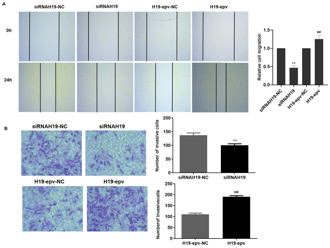
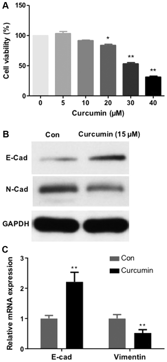
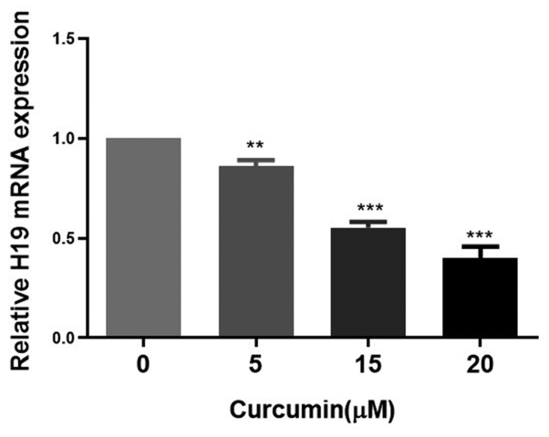
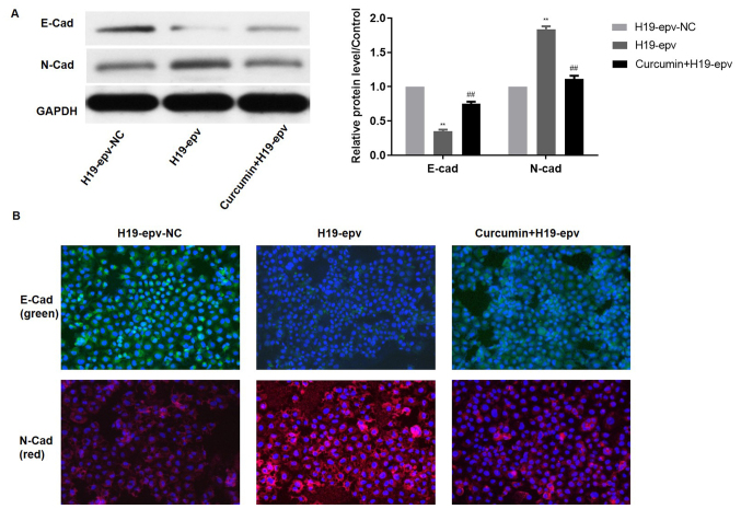
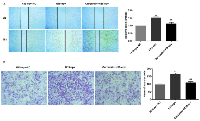
The H19 long non‑coding RNA is involved in the development of tamoxifen resistance in breast cancer. However, the relationship between H19 and the metastatic potential and treatment options for tamoxifen‑resistant (TAMR) breast cancer is not completely understood. Curcumin inhibits cellular proliferation, migration and invasiveness in several cancer types, including pancreatic cancer, breast cancer and chronic myeloid leukemia. The present study aimed to investigate the role of H19 in MCF‑7/TAMR cell epithelial‑mesenchymal transition (EMT), migration and invasiveness, and to assess the ability of curcumin to inhibit H19‑mediated effects. Reverse transcription‑quantitative PCR and western blot analysis were conducted to detect the gene or protein expression. Cell Counting Kit‑8, wound healing and Transwell invasion assays were performed to estimate the capabilities of cell viability, invasion and migration. H19 overexpression enhanced MCF‑7/TAMR cell EMT, invasion and migration by upregulating Snail. Furthermore, curcumin notably decreased the expression levels of epithelial marker E‑cadherin and markedly increased the expression levels of mesenchymal marker N‑cadherin in MCF‑7/TAMR cells compared with the control group. In addition, following treatment with curcumin for 48 h, H19 expression was decreased in a dose‑dependent manner. Moreover, curcumin treatment for 48 h significantly attenuated H19‑induced alterations in N‑cadherin and E‑cadherin expression levels. Curcumin also prevented H19‑induced invasion and migration. The present study indicated that H19 may serve as a promoting factor of EMT, invasion and migration in MCF‑7/TAMR cells, suggesting that curcumin may prevent H19‑associated metastasis. Therefore, curcumin may serve as a promising therapeutic drug for patients with TAMR breast cancer.
Keywords: tamoxifen‑resistance; epithelial‑mesenchymal transition; breast cancer; H19 imprinted maternally expressed transcript; curcumin.
Figures


References
-
- Poggio F, Ceppi M, Lambertini M, Bruzzi P, Ugolini D, Bighin C, Levaggi A, Giraudi S, D'Alonzo A, Vaglica M, et al. Concurrent versus sequential adjuvant chemo-endocrine therapy in hormone-receptor positive early stage breast cancer patients: A systematic review and meta-analysis. Breast. 2017;33:104–108. doi: 10.1016/j.breast.2017.03.011. - DOI - PubMed
-
- Zhang Y, Liu Y. Endocrine therapy for breast cancer: Past and present. Zhonghua Yi Shi Za Zhi. 2015;45:28–32. (In Chinese) - PubMed
-
- Early Breast Cancer Trialists' Collaborative Group (EBCTCG) Davies C, Godwin J, Gray R, Clarke M, Cutter D, Darby S, McGale P, Pan HC, Taylor C, et al. Relevance of breast cancer hormone receptors and other factors to the efficacy of adjuvant tamoxifen: patient-level metaanalysis of randomised trials. Lancet. 2011;378:771–784. doi: 10.1016/S0140-6736(11)60993-8. - DOI - PMC - PubMed
-
- Burstein HJ, Temin S, Anderson H, Buchholz TA, Davidson NE, Gelmon KE, Giordano SH, Hudis CA, Rowden D, Solky AJ, et al. Adjuvant endocrine therapy for women with hormone receptor-positive breast cancer: American society of clinical oncology clinical practice guideline focused update. J Clin Oncol. 2014;32:2255–2269. doi: 10.1200/JCO.2013.54.2258. - DOI - PMC - PubMed
MeSH terms
Substances
LinkOut - more resources
Full Text Sources
Medical
Research Materials
